College Physics (2012) by Manjula Sharma, Paul Peter Urone, et al - HTML preview
Download the book in PDF, ePub, Kindle for a complete version.
⎛
⎞2
(17.39)
a = ⎝ Z 2 − Z 1⎠
⎛
⎞2,
⎝ Z 1 + Z 2⎠
where Z 1 and Z 2 are the acoustic impedances of the two media making up the boundary. A reflection coefficient of zero (corresponding to total
transmission and no reflection) occurs when the acoustic impedances of the two media are the same. An impedance “match” (no reflection) provides
an efficient coupling of sound energy from one medium to another. The image formed in an ultrasound is made by tracking reflections (as shown in
Figure 17.44) and mapping the intensity of the reflected sound waves in a two-dimensional plane.
Example 17.7 Calculate Acoustic Impedance and Intensity Reflection Coefficient: Ultrasound and Fat Tissue
(a) Using the values for density and the speed of ultrasound given in Table 17.5, show that the acoustic impedance of fat tissue is indeed
1.34×10⁶ kg/(m2 ·s) .
(b) Calculate the intensity reflection coefficient of ultrasound when going from fat to muscle tissue.
Strategy for (a)
The acoustic impedance can be calculated using Z = ρv and the values for ρ and v found in Table 17.5.
Solution for (a)
(1) Substitute known values from Table 17.5 into Z = ρv .
(17.40)
Z = ρv = ⎛⎝925 kg/m3⎞⎠(1450 m/s)
616 CHAPTER 17 | PHYSICS OF HEARING
(2) Calculate to find the acoustic impedance of fat tissue.
(17.41)
1.34×10⁶ kg/(m2 ·s)
This value is the same as the value given for the acoustic impedance of fat tissue.
Strategy for (b)
⎛
⎞2
The intensity reflection coefficient for any boundary between two media is given by a = ⎝ Z 2 − Z 1⎠
⎛
⎞2 , and the acoustic impedance of muscle is
⎝ Z 1 + Z 2⎠
given in Table 17.5.
Solution for (b)
⎛
⎞2
Substitute known values into a = ⎝ Z 2 − Z 1⎠
⎛
⎞2 to find the intensity reflection coefficient:
⎝ Z 1 + Z 2⎠
2
(17.42)
⎛
⎞2
⎛
a = ⎝ Z 2 − Z 1⎠
⎝1.34×10⁶ kg/(m2 · s) − 1.70×10⁶ kg/(m2 · s)⎞⎠
⎛
⎞2 =
2 = 0.014
⎝ Z 1 + Z 2⎠
⎛⎝1.70×10⁶ kg/(m2· s) + 1.34×10⁶ kg/(m2· s)⎞⎠
Discussion
This result means that only 1.4% of the incident intensity is reflected, with the remaining being transmitted.
The applications of ultrasound in medical diagnostics have produced untold benefits with no known risks. Diagnostic intensities are too low (about
10−2 W/m2 ) to cause thermal damage. More significantly, ultrasound has been in use for several decades and detailed follow-up studies do not
show evidence of ill effects, quite unlike the case for x-rays.
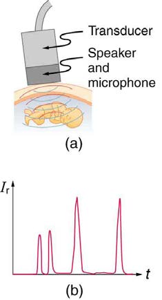
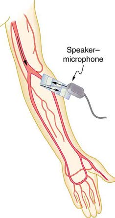
Figure 17.44 (a) An ultrasound speaker doubles as a microphone. Brief bleeps are broadcast, and echoes are recorded from various depths. (b) Graph of echo intensity
versus time. The time for echoes to return is directly proportional to the distance of the reflector, yielding this information noninvasively.
The most common ultrasound applications produce an image like that shown in Figure 17.45. The speaker-microphone broadcasts a directional
beam, sweeping the beam across the area of interest. This is accomplished by having multiple ultrasound sources in the probe’s head, which are
phased to interfere constructively in a given, adjustable direction. Echoes are measured as a function of position as well as depth. A computer
constructs an image that reveals the shape and density of internal structures.
CHAPTER 17 | PHYSICS OF HEARING 617
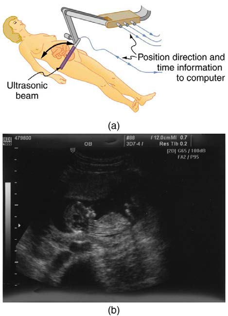
Figure 17.45 (a) An ultrasonic image is produced by sweeping the ultrasonic beam across the area of interest, in this case the woman’s abdomen. Data are recorded and
analyzed in a computer, providing a two-dimensional image. (b) Ultrasound image of 12-week-old fetus. (credit: Margaret W. Carruthers, Flickr)
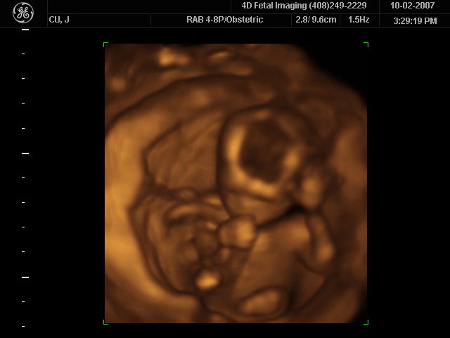
How much detail can ultrasound reveal? The image in Figure 17.45 is typical of low-cost systems, but that in Figure 17.46 shows the remarkable detail possible with more advanced systems, including 3D imaging. Ultrasound today is commonly used in prenatal care. Such imaging can be used
to see if the fetus is developing at a normal rate, and help in the determination of serious problems early in the pregnancy. Ultrasound is also in wide
use to image the chambers of the heart and the flow of blood within the beating heart, using the Doppler effect (echocardiology).
Whenever a wave is used as a probe, it is very difficult to detect details smaller than its wavelength λ . Indeed, current technology cannot do quite
this well. Abdominal scans may use a 7-MHz frequency, and the speed of sound in tissue is about 1540 m/s—so the wavelength limit to detail would
be λ = v w
f = 1540 m/s = 0.22 mm . In practice, 1-mm detail is attainable, which is sufficient for many purposes. Higher-frequency ultrasound
7×10⁶ Hz
would allow greater detail, but it does not penetrate as well as lower frequencies do. The accepted rule of thumb is that you can effectively scan to a
depth of about 500 λ into tissue. For 7 MHz, this penetration limit is 500×0.22 mm , which is 0.11 m. Higher frequencies may be employed in
smaller organs, such as the eye, but are not practical for looking deep into the body.
Figure 17.46 A 3D ultrasound image of a fetus. As well as for the detection of any abnormalities, such scans have also been shown to be useful for strengthening the
emotional bonding between parents and their unborn child. (credit: Jennie Cu, Wikimedia Commons)
In addition to shape information, ultrasonic scans can produce density information superior to that found in X-rays, because the intensity of a reflected
sound is related to changes in density. Sound is most strongly reflected at places where density changes are greatest.
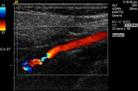
Another major use of ultrasound in medical diagnostics is to detect motion and determine velocity through the Doppler shift of an echo, known as
Doppler-shifted ultrasound. This technique is used to monitor fetal heartbeat, measure blood velocity, and detect occlusions in blood vessels, for
example. (See Figure 17.47.) The magnitude of the Doppler shift in an echo is directly proportional to the velocity of whatever reflects the sound.
618 CHAPTER 17 | PHYSICS OF HEARING
Because an echo is involved, there is actually a double shift. The first occurs because the reflector (say a fetal heart) is a moving observer and
receives a Doppler-shifted frequency. The reflector then acts as a moving source, producing a second Doppler shift.
Figure 17.47 This Doppler-shifted ultrasonic image of a partially occluded artery uses color to indicate velocity. The highest velocities are in red, while the lowest are blue. The blood must move faster through the constriction to carry the same flow. (credit: Arning C, Grzyska U, Wikimedia Commons)
A clever technique is used to measure the Doppler shift in an echo. The frequency of the echoed sound is superimposed on the broadcast frequency,
producing beats. The beat frequency is F B = ∣ f 1 − f 2 ∣ , and so it is directly proportional to the Doppler shift ( f 1 − f 2 ) and hence, the reflector’s velocity. The advantage in this technique is that the Doppler shift is small (because the reflector’s velocity is small), so that great accuracy
would be needed to measure the shift directly. But measuring the beat frequency is easy, and it is not affected if the broadcast frequency varies
somewhat. Furthermore, the beat frequency is in the audible range and can be amplified for audio feedback to the medical observer.
Uses for Doppler-Shifted Radar
Doppler-shifted radar echoes are used to measure wind velocities in storms as well as aircraft and automobile speeds. The principle is the same
as for Doppler-shifted ultrasound. There is evidence that bats and dolphins may also sense the velocity of an object (such as prey) reflecting
their ultrasound signals by observing its Doppler shift.
Example 17.8 Calculate Velocity of Blood: Doppler-Shifted Ultrasound
Ultrasound that has a frequency of 2.50 MHz is sent toward blood in an artery that is moving toward the source at 20.0 cm/s, as illustrated in
figures.)
a. What frequency does the blood receive?
b. What frequency returns to the source?
c. What beat frequency is produced if the source and returning frequencies are mixed?
Figure 17.48 Ultrasound is partly reflected by blood cells and plasma back toward the speaker-microphone. Because the cells are moving, two Doppler shifts are
produced—one for blood as a moving observer, and the other for the reflected sound coming from a moving source. The magnitude of the shift is directly proportional to
blood velocity.
Strategy
⎛
v
⎞
⎛ v
⎞
The first two questions can be answered using f
w
w ± v obs
obs = f s⎝ v w ± v s⎠ and f obs = f s⎝
v w
⎠ for the Doppler shift. The last question
asks for beat frequency, which is the difference between the original and returning frequencies.
CHAPTER 17 | PHYSICS OF HEARING 619
Solution for (a)
(1) Identify knowns:
• The blood is a moving observer, and so the frequency it receives is given by
⎛ v
⎞
(17.43)
f
w ± v obs
obs = f s⎝
v w ⎠.
•
v b is the blood velocity ( v obs here) and the plus sign is chosen because the motion is toward the source.
(2) Enter the given values into the equation.
⎞
(17.44)
f
1540 m/s+0.2 m/s
obs = (2,500 , 000 Hz)⎛⎝
1540 m/s
⎠
(3) Calculate to find the frequency: 20,500,325 Hz.
Solution for (b)
(1) Identify knowns:
• The blood acts as a moving source.
• The microphone acts as a stationary observer.
• The frequency leaving the blood is 2,500,325 Hz, but it is shifted upward as given by
⎛
v
⎞
(17.45)
f
w
obs = f s⎝ v w – v b⎠.
f obs is the frequency received by the speaker-microphone.
• The source velocity is v b .
• The minus sign is used because the motion is toward the observer.
The minus sign is used because the motion is toward the observer.
(2) Enter the given values into the equation:
⎞
(17.46)
f
1540 m/s
obs = (2,500 , 325 Hz)⎛⎝1540 m/s − 0.200 m/s⎠
(3) Calculate to find the frequency returning to the source: 2,500,649 Hz.
Solution for (c)
(1) Identify knowns:
• The beat frequency is simply the absolute value of the difference between f s and f obs , as stated in:
f
(17.47)
B = ∣ f obs − f s ∣ .
(2) Substitute known values:
(17.48)
∣ 2,500 , 649 Hz − 2,500 , 000 Hz ∣
(3) Calculate to find the beat frequency: 649 Hz.
Discussion
The Doppler shifts are quite small compared with the original frequency of 2.50 MHz. It is far easier to measure the beat frequency than it is to
measure the echo frequency with an accuracy great enough to see shifts of a few hundred hertz out of a couple of megahertz. Furthermore,
variations in the source frequency do not greatly affect the beat frequency, because both f s and f obs would increase or decrease. Those
changes subtract out in f B = ∣ f obs − f s ∣ .
Industrial and Other Applications of Ultrasound
Industrial, retail, and research applications of ultrasound are common. A few are discussed here. Ultrasonic cleaners have many uses. Jewelry,
machined parts, and other objects that have odd shapes and crevices are immersed in a cleaning fluid that is agitated with ultrasound typically
about 40 kHz in frequency. The intensity is great enough to cause cavitation, which is responsible for most of the cleansing action. Because
cavitation-produced shock pressures are large and well transmitted in a fluid, they reach into small crevices where even a low-surface-tension
cleaning fluid might not penetrate.
Sonar is a familiar application of ultrasound. Sonar typically employs ultrasonic frequencies in the range from 30.0 to 100 kHz. Bats, dolphins,
submarines, and even some birds use ultrasonic sonar. Echoes are analyzed to give distance and size information both for guidance and finding
prey. In most sonar applications, the sound reflects quite well because the objects of interest have significantly different density than the medium
in which they travel. When the Doppler shift is observed, velocity information can also be obtained. Submarine sonar can be used to obtain such
information, and there is evidence that some bats also sense velocity from their echoes.
Similarly, there are a range of relatively inexpensive devices that measure distance by timing ultrasonic echoes. Many cameras, for example, use
such information to focus automatically. Some doors open when their ultrasonic ranging devices detect a nearby object, and certain home
security lights turn on when their ultrasonic rangers observe motion. Ultrasonic “measuring tapes” also exist to measure such things as room
dimensions. Sinks in public restrooms are sometimes automated with ultrasound devices to turn faucets on and off when people wash their
hands. These devices reduce the spread of germs and can conserve water.
620 CHAPTER 17 | PHYSICS OF HEARING
Ultrasound is used for nondestructive testing in industry and by the military. Because ultrasound reflects well from any large change in density, it
can reveal cracks and voids in solids, such as aircraft wings, that are too small to be seen with x-rays. For similar reasons, ultrasound is also
good for measuring the thickness of coatings, particularly where there are several layers involved.
Basic research in solid state physics employs ultrasound. Its attenuation is related to a number of physical characteristics, making it a useful
probe. Among these characteristics are structural changes such as those found in liquid crystals, the transition of a material to a superconducting
phase, as well as density and other properties.
These examples of the uses of ultrasound are meant to whet the appetites of the curious, as well as to illustrate the underlying physics of
ultrasound. There are many more applications, as you can easily discover for yourself.
Why is it possible to use ultrasound both to observe a fetus in the womb and also to destroy cancerous tumors in the body?
Solution
Ultrasound can be used medically at different intensities. Lower intensities do not cause damage and are used for medical imaging. Higher
intensities can pulverize and destroy targeted substances in the body, such as tumors.
Glossary
acoustic impedance: property of medium that makes the propagation of sound waves more difficult
antinode: point of maximum displacement
bow wake: V-shaped disturbance created when the wave source moves faster than the wave propagation speed
Doppler effect: an alteration in the observed frequency of a sound due to motion of either the source or the observer
Doppler shift: the actual change in frequency due to relative motion of source and observer
Doppler-shifted ultrasound: a medical technique to detect motion and determine velocity through the Doppler shift of an echo
fundamental: the lowest-frequency resonance
harmonics: the term used to refer collectively to the fundamental and its overtones
hearing: the perception of sound
infrasound: sounds below 20 Hz
intensity reflection coefficient: a measure of the ratio of the intensity of the wave reflected off a boundary between two media relative to the
intensity of the incident wave
intensity: the power per unit area carried by a wave
loudness: the perception of sound intensity
node: point of zero displacement
note: basic unit of music with specific names, combined to generate tunes
overtones: all resonant frequencies higher than the fundamental
phon: the numerical unit of loudness
pitch: the perception of the frequency of a sound
sonic boom: a constructive interference of sound created by an object moving faster than sound
sound intensity level: a unitless quantity telling you the level of the sound relative to a fixed standard
sound pressure level: the ratio of the pressure amplitude to a reference pressure
sound: a disturbance of matter that is transmitted from its source outward
timbre: number and relative intensity of multiple sound frequencies
tone: number and relative intensity of multiple sound frequencies
ultrasound: sounds above 20,000 Hz
Section Summary
• Sound is a disturbance of matter that is transmitted from its source outward.
CHAPTER 17 | PHYSICS OF HEARING 621
• Sound is one type of wave.
• Hearing is the perception of sound.
17.2 Speed of Sound, Frequency, and Wavelength
The relationship of the speed of sound v w , its frequency f , and its wavelength λ is given by
v w = fλ,
which is the same relationship given for all waves.
In air, the speed of sound is related to air temperature T by
v w = (331 m/s) T
273 K.
v w is the same for all frequencies and wavelengths.
17.3 Sound Intensity and Sound Level
• Intensity is the same for a sound wave as was defined for all waves; it is
I = PA,
where P is the power crossing area A . The SI unit for I is watts per meter squared. The intensity of a sound wave is also related to the
pressure amplitude Δ p
⎛
2
I = ⎝Δ p⎞⎠
2 ρv ,
w
where ρ is the density of the medium in which the sound wave travels and v w is the speed of sound in the medium.
• Sound intensity level in units of decibels (dB) is
β (dB) = 10 log ⎛ I ⎞
10⎝ I 0⎠,
where I 0 = 10–12 W/m2 is the threshold intensity of hearing.
17.4 Doppler Effect and Sonic Booms
• The Doppler effect is an alteration in the observed frequency of a sound due to motion of either the source or the observer.
• The actual change in frequency is called the Doppler shift.
• A sonic boom is constructive interference of sound created by an object moving faster than sound.
• A sonic boom is a type of bow wake created when any wave source moves faster than the wave propagation speed.
• For a stationary observer and a moving source, the observed frequency f obs is:
f
⎛ v w ⎞
obs = f s⎝ v w ± v s⎠,
where f s is the frequency of the source, v s is the speed of the source, and v w is the speed of sound. The minus sign is used for motion
toward the observer and the plus sign for motion away.
• For a stationary source and moving observer, the observed frequency is:
f
⎛ v w ± v obs⎞
obs = f s⎝
v w ⎠,
where v obs is the speed of the observer.
17.5 Sound Interference and Resonance: Standing Waves in Air Columns
• Sound interference and resonance have the same properties as defined for all waves.
• In air columns, the lowest-frequency resonance is called the fundamental, whereas all higher resonant frequencies are called overtones.
Collectively, they are called harmonics.
• The resonant frequencies of a tube closed at one end are:
fn = nv w
4 L, n = 1, 3, 5...,
f 1 is the fundamental and L is the length of the tube.
• The resonant frequencies of a tube open at both ends are:
fn = nv w
2 L, n = 1, 2, 3...
• The range of audible frequencies is 20 to 20,000 Hz.
• Those sounds above 20,000 Hz are ultrasound, whereas those below 20 Hz are infrasound.
• The perception of frequency is pitch.
• The perception of intensity is loudness.
• Loudness has units of phons.
622 CHAPTER 17 | PHYSICS OF HEARING
• The acoustic impedance is defined as:
Z = ρv,
ρ is the density of a medium through which the sound travels and v is the speed of sound through that medium.