

Endocrine Physiology: 100-Questions and Explanatory Answers for Students by Dr. Mustafa Fadil Mohammed - HTML preview
Download the book in PDF, ePub, Kindle for a complete version.
Endocrine Physiology: 100 Questions and
Explanatory Answers for Students
Mustafa Fadil Mohammed
PhD/Physiology
Management and Science University
Malaysia
This book is intended to review, assess, evaluate and further improve the student’s knowledge in the endocrine physiology that presented in an easy to understand, graphical and tubulated structure format.
This book's multiple-choice questions have been extensively prepared to emphasize the relative significance of the various endocrine glands and to act as a foundational source for endocrine physiology.
The aim of this book is to provide the learner with an in-depth, rapid review of hormone functions and the effects of abnormal hormone levels. It also functions as an essential reading for the endocrine physiology course.
The reader is encouraged to send comments, critiques and recommendations to improve subsequent editions.
Mustafa Fadil Mohammed
Chapter1: Introduction to the endocrine system
1
Chapter2: Hormone receptors
10
Chapter3: Hypothalamus and posterior pituitary hormones
16
Chapter4: Anterior pituitary – Growth hormone and prolactin 23
Chapter5: Adrenal glands
29
Chapter6: Thyroid gland
35
Chapter7: Reproductive hormones
42
Chapter8: Pancreas
49
Chapter9: Parathyroid gland
55
Chapter10: Miscellaneous hormones
62
73
__________________________________________________
CHAPTER 1: INTRODUCTION TO THE ENDOCRINE SYSTEM
1.
Hormones are usually classified based on their chemical structure. Aldosterone would be categorized as _____.
A.
protein hormone
B.
steroid hormone
C.
amines and amino acid hormone
D.
peptides and polypeptides hormone
2.
Regarding endocrine glands, which of the following is FALSE?
A.
They produce hormonal substances.
B.
They have ducts.
C.
Adrenal gland is an example of the endocrine gland.
D.
They have no ducts.
3.
Thyroid stimulating hormone (TSH) induces thyroid hormone production. TSH is then inhibited by thyroid hormone. Which of the following scenarios describe this relationship?
A.
Membrane receptor transformation.
B.
Non-hormonal chemical messengers.
C.
Negative feedback regulation.
D.
Intercellular communication.
4.
Which of the following unsaturated fatty acid is the source of eicosanoids' synthesis?
A.
Oleic acid
B.
Linoleic acid
C.
Linolenic acid
D.
Arachidonic acid
5.
Select the TRUE statement regarding the half-life of a hormone: A.
A hormone is half the time taken for it to disappear from the blood.
B.
Insulin is between five and ten hours.
C.
Thyroxine is longer than that of adrenaline.
D.
Aldosterone is longer than cortisol.
6.
Which endocrine glands located anterior to the neck?
A.
Thyroid glands.
B.
Pancreas.
C.
Adrenal glands.
D.
Pineal gland.
7.
In an examination room, a student has an elevated heart rate and increased breathing rate. What type of endocrine system stimulus did the student receive?
A.
Hormonal.
B.
Negative feedback.
C.
Neural.
D.
Positive feedback.
8.
The endocrine system works together with the ________ system to maintain the body’s homeostasis?
A.
Digestive system.
B.
Nervous system.
C.
Respiratory system.
D.
Reproductive system.
The largest endocrine gland(s) that makes 3 hormones that affect the metabolism is the _____.
A.
pancreas
B.
adrenal glands
C.
thyroid gland
D.
pituitary gland
10.
Which of the following hormones is stored in secretory vesicles prior to secretion?
A.
Estrogen.
B.
Progesterone.
C.
Aldosterone
D.
Insulin
1.
(B) Aldosterone is a steroid hormone. Hormones can be classified into three types.
Proteins, steroid hormones and amine hormones.
Classification of hormones according to their chemical structures.
Classes of hormones
Characteristics
Examples
Protein/
Peptide 1. Made of chains of amino acids. Protein hormones: hormones
2. These hormones are stored in Growth
hormone
(GH),
secretory vesicles.
Prolactin (PRL), Parathyroid
3. Cell membrane receptor.
hormone (PTH).
4. Water soluble.
5. Do not require carrier protein.
Peptide hormones:
Antidiuretic hormone (ADH)
Oxytocin and Somatostatin
(SS).
Steroid hormones
1. Derived from Cholesterol.
Adrenal cortex hormones:
2. Lipid soluble.
Aldosterone and Cortisol.
3. Immediately released from the Reproductive hormones: cell following synthesis
Estrogen, Testosterone and
4.
Cytoplasmic
or
nuclear Progesterone.
receptors.
5. Require carrier proteins.
Amine hormones
1. Derived from a single amino Adrenal medulla: acid for example tyrosine.
Norepinephrine
(NE)
and
2. Receptors either on:
Epinephrine (Epi).
Cell surface (Catecholamines)
Thyroid hormones:
Intracellular (Thyroid hormone). Thyroxine (T4)
and
Triiodothyronine (T3).
2.
(B) Endocrine glands are a ductless gland made up epithelial cells that release chemical signals called hormones. Hormones pass through the bloodstream to react on target cells receptors, influencing the cell’s metabolic activities.
Hormones of the hypothalamus
Hormones
Major actions
Thyrotropin releasing hormone:
Stimulate release of TSH
(TRH)
and prolactin secretion
Gonadotropin releasing
Stimulate release of LH and FSH
hormone (GnRH)
Corticotropin releasing hormone
Stimulate release of ACTH
(CRH)
Growth hormone-releasing
Stimulate secretion of growth hormone (GH)
hormone (GHRH)
Growth hormone-inhibitory
Inhibit the release of GH
hormone (GHIH) also known as
somatostatin
Prolactin-inhibitory factor (PIF)
Inhibit release of Prolactin
also known as dopamine
Hormones of the pituitary gland Pituitary
Hormones
Major actions
gland
Anterior
Adrenocorticotropic
Stimulate steroid hormones formation from the pituitary
hormone (ACTH)
adrenal
cortex
(aldosterone,
cortisol
and
androgens).
Growth
hormone Promote release of somatomedins from the liver.
(GH)
Stimulate bone growth.
Increase blood glucose and fatty acids.
luteinizing hormone In males stimulate the release of testosterone (LH)
from the testes Leydig cells.
In females stimulate ovulation and the release of progesterone.
follicle
stimulating In males stimulate sperm maturation.
hormone (FSH)
In females stimulate ovarian follicle development.
Prolactin
Stimulate milk production and growth of breast.
Thyroid
stimulating Stimulate the synthesis and secretion of thyroid hormone (TSH)
hormones T3 and T4
Posterior
Antidiuretic hormone Maintain plasma osmolality by promoting water pituitary
(ADH) also known reabsorption from the nephron.
as
arginine Arteriolar vasoconstriction.
vasopressin
Stimulate the release of ACTH
Oxytocin
Stimulate uterine contraction during labor
Stimulate milk ejection
Hormones of the adrenal glands
Adrenal
Hormones
Major actions
gland
Adrenal
Aldosterone
Maintain blood volume by promoting Na+ and water cortex
reabsorption from the kidney nephrons.
Stimulate urinary excretion of K+ and H+
Cortisol
Increase plasma glucose concentration.
Anti-inflammatory actions.
Androgens
In females stimulate growth of pubic and axillary hair and increase libido.
Adrenal
Epinephrine and Promote fight or flight responses as the sympathetic medulla
norepinephrine
nervous system
Hormones of thyroid and parathyroid glands
Hormones
Major actions
Thyroid
Triiodothyronine (T3) Increase basal metabolic rate and oxygen and Thyroxine (T4)
consumption.
Enhance neural and bone growth and development.
Calcitonin
Decreases serum Ca++
Parathyroid Parathyroid hormone Increases serum Ca++ levels by resorption of Ca++
(PTH)
from bone and reabsorption of Ca++ from the distal tubule.
Pancreas
Hormones
Major actions
Pancreas
Insulin
Promote storage of glucose
Promote storage of triglyceride
Glucagon
Increases blood glucose
Hormones of the gonads
Gonads
Hormones
Major actions
Testes
Testosterone
Maintenance of spermatogenesis
Maintenance of secondary sex characteristic including growth of facial, pubic and axillary hair.
Maintenance of muscle mass
Ovary
Estrogens
Maintenance of secondary sex characteristic
including breast development.
Stimulate cervical mucus production.
Stimulate endometrial cells proliferation.
Increase libido
Progesterone
Maintenance of pregnancy.
Maintain the secretion of the endometrium during the secretory phase of menstrual cycle.
In addition, a number of organs including the gastrointestinal tract, kidney, liver heart, skin, adipose tissue can be regarded as an endocrine gland because they can all release hormones but they do not form a distinct endocrine gland.
Other endocrine organs
Organs
Hormones
Major actions
Heart
Atrial natriuretic peptide (ANP)
Maintain blood volume by promoting
excretion of Na+ and water.
Kidney
Renin
Maintain
arterial
blood
pressure
by
activating the renin angiotensin aldosterone
system.
1-25-Dihydroxycholecalciferol
Stimulate absorption of calcium and
phosphate from the gastrointestinal tract.
Erythropoietin
Stimulate red blood cell production.
GIT
Gastrin
Stimulate HCl production by the parietal
cells of gastric mucosa.
Cholecystokinin (CCK)
Stimulate
the
release
of
pancreatic
enzymes.
Inhibits stomach motility.
Secretin
Increases
HCO -
3
secretion
by
the
pancreatic duct.
Placenta Human chorionic gonadotropin Prevent the regression of corpus luteum (hCG)
and maintain pregnancy.
Human Chorionic
Stimulate
breast
development
during
Somatomammotropin (hCS)
pregnancy
3.
(C) Negative feedback prevents over secretion of the hormone or over activity at the target tissue such as regulation of thyroid hormone by thyroid stimulating hormone (TSH).
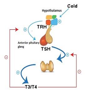
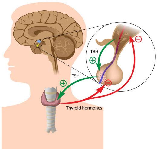
Negative Feedback Mechanism: A stimulus such as cold causes an increase in thyrotropin-releasing hormone (TRH) from the hypothalamus, which in turn acts to increase the secretion of thyroid-stimulating hormone (TSH) from the anterior pituitary. The high levels of TSH stimulate the thyroid gland to produce thyroid hormones (T3 and T4). One of the effects of thyroid hormones is to act on both the hypothalamus and anterior pituitary to inhibit the production of TRH and TSH, respectively. Thus, the blood levels of TRH and TSH are maintained by a negative feedback system to prevent over secretion of these hormones caused by the stimulus in this case cold.
Positive feedback is two or more variables, if one increases the second one, the second one, in turn, increases the first one such as estrogen during ovulation.
Cyclical variations occur in hormone release such as growth hormone.
4.
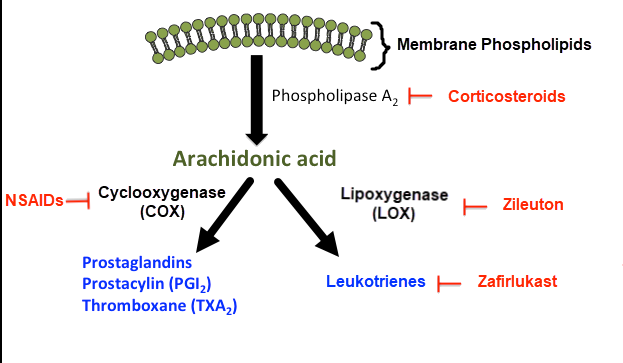
(D) Arachidonic acid is the precursor of eicosanoids synthesis. Arachidonic acid is derived from the membrane phospholipids when the enzyme phospholipase A2 is activated via G protein (Gq). Eicosanoids which include prostaglandins, thromboxanes, prostacyclins and leukotrienes are locally produced regulatory hormones. The synthesis of eicosanoids is differed according to the enzymes that expressed in the target cell (cyclooxygenase or lipoxygenase).
Arachidonic acid, which is produced from the phospholipid in the cell membrane, is the precursor of eicosanoids. Phospholipids in cell membranes are converted into arachidonic acid by the enzyme phospholipase A2. Two distinct processes the cyclooxygenase and lipoxygenase convert arachidonic acid into prostaglandins, thromboxanes, prostacyclins and leukotrienes.
5.
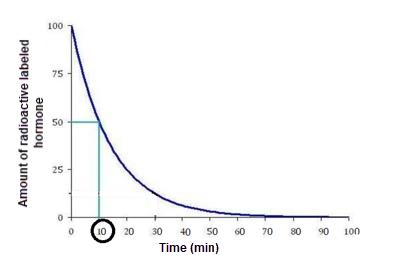
(C) The half-life is the time taken to reduce hormone concentration (due to metabolism and excretion) by one half. The half-life of protein bounds hormones such as cortisol, thyroid hormones, testosterone is longer than those which does not bound to plasma proteins such as most peptide hormones and aldosterone.
Half-life is the time interval between 0 and half the concentration of the hormone. In this example, the plasma half-life of a hormone is 10min. Normally peptide hormones have a short half-life as compared to steroid hormones.
6.
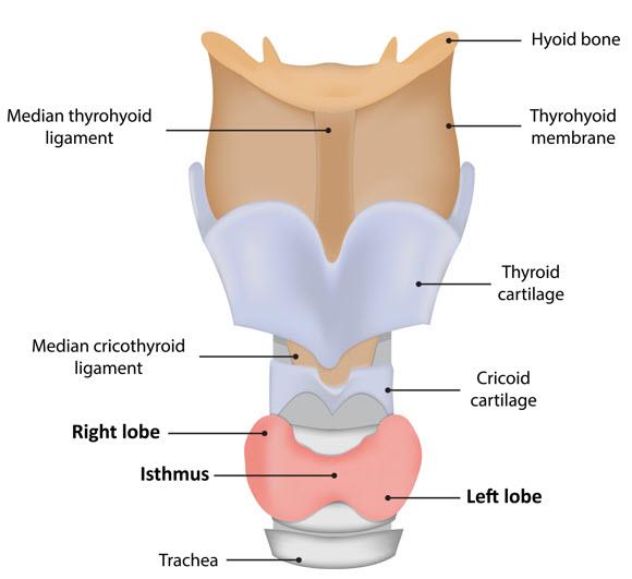
(A) The thyroid gland is the largest endocrine gland, located in the anterior neck, consists of two lateral lobes connected by a median tissue mass called the isthmus.
Composed of follicles that produce the glycoprotein thyroglobulin.
The major endocrine glands and their location Glands
Location
Hypothalamus
Central nervous system (brain)
Pituitary gland
Central nervous system
Pineal gland
Central nervous system (brain)
Thyroid gland
Neck (Anterior to trachea)
Parathyroid gland
Neck (Posterior to thyroid gland)
Adrenal glands
Abdominal cavity (above the kidney)
Pancreas
Abdominal cavity
Testes
Within the scrotum
Ovaries
Pelvic cavity
7.
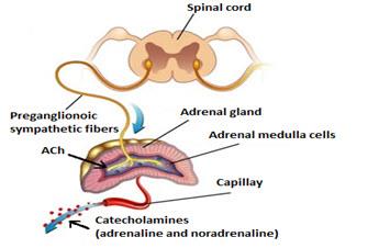
(C) Activation of preganglionic sympathetic nervous system (SNS) fibers directly stimulate the adrenal medulla gland to release epinephrine and norepinephrine.
Negative feedback is to maintain a ‘set point’ of hormone levels by a correction mechanism that opposes (negative) the changes from this point. Hormonal stimuli refer to the release of hormones in response to hormones produced by other endocrine organs. For example, the release of thyroid hormones is controlled by the anterior pituitary, thyroid-stimulating hormone (TSH) hormone which is in turn controlled by hypothalamic releasing factors (TRH).
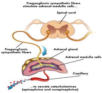
Release of adrenal medulla hormones in response to neural stimuli by sympathetic nervous system
8.
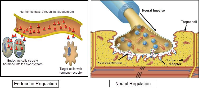
(B) Both the endocrine and the nervous system act to maintain homeostasis through the production of hormones and neurotransmitters respectively.
Endocrine system vs Nervous system
Comparison between nervous and the endocrine system Feature
Endocrine system
Nervous system
Overall function
Maintain homeostasis
Maintain homeostasis
Control
by
feedback By negative and positive By nervous reflexes mechanism
feedback
Effector cells
Target cells throughout that Postsynaptic cells the body have
receptors
for
the in muscle, neurons and
specific hormone
glandular tissue
Chemical messenger
Hormone
Neurotransmitter
Cells
that
secrete
the Glandular epithelial cells or Neurons chemical messenger
neurosecretory cells
Distance travel of chemical Long (through bloodstream) Short (though synapse)
messenger
Location of receptors
On the plasma or within cell On the plasma membrane membrane
Speed of communication
Slow
Fast
9.
(C) Thyroid gland is the largest endocrine gland, located in the anterior neck, consists of two lateral lobes connected by a median tissue mass called the isthmus.
Location of thyroid hormone
10.
(D) Endocrine cells that secrete peptides store hormones in secretory vesicles and release them via exocytosis when a stimulus initiates hormone secretion. This is true for insulin, which is produced in the β cells of pancreas. In contrast, steroid hormones are not stored in secretory vesicles and released rapidly once synthesized.
Comparison between peptides and steroids hormones Characteristic
Peptides
Steroids
Source of hormone
Amino acids
Cholesterol
Storage of hormones
Secretory vesicles
Stored as precursor
Location of receptors
Cell surface membrane
Intracellular (cytoplasm or
nucleus)
Transport
with
carrier No
Yes
protein
1.
Action of hormone, on the target cells, depending on binding of hormone with A.
hormone
B.
receptor
C.
steroids
D.
proteins
2.
Receptors for peptide hormones are located ____________.
A.
on the cell membrane
B.
in the cytoplasm
C.
in the ribosome
D.
in the nucleus
3.
Which of the following signaling molecules DO NOT couple to G proteins?
A.
Cyclic adenosine monophosphate (cAMP).
B.
Diacylglycerol (DAG).
C.
Inositol triphosphate (IP3).
D.
Tyrosine kinase.
4.
A newly developed drug has been observed to bind to an intracellular hormone receptor. If ingested, residue from this drug could disrupt levels of
.
A.
melatonin
B.
thyroid hormone
C.
growth hormone
D.
insulin
5.
Which one of the following is NOT typical of the cellular responses that occurs after a hormone binds to its target cells:
A.
plasma membrane permeability changes
B.
cellular mutations occur
C.
enzymes are activated or inactivated
D.
mitosis is stimulated
6.
Being lipid soluble, steroids hormone can cause all the following cellular activities EXCEPT:
A.
catalyze cyclic AMP
B.
diffuse through the plasma membranes of target cells C.
enter the nucleus
D.
activate genes to transcribe mRNA for protein synthesis 7.
Which of the following hormones activate enzyme-linked receptors?
A.
Antidiuretic hormone
B.
Growth hormone
C.
Cortisol
D.
Aldosterone
8.
Atrial natriuretic peptide increases the formation of A.
cyclic GMP
B.
tyrosine kinase
C.
cyclic AMP
D.
diacylglycerol
All of the following bind to intracellular receptors EXCEPT: A.
thyroxine
B.
aldosterone
C.
1, 25-dihydroxycholecalciferol
D.
parathyroid hormone
10.
All the following second messenger is increased when antidiuretic hormone (ADH) acts on its receptors EXCEPT:
A.
cyclic AMP (cAMP)
B.
tyrosine kinase (TK)
C.
diacylglycerol (DAG)
D.
inositol triphosphate (IP3)
Answers:
1.
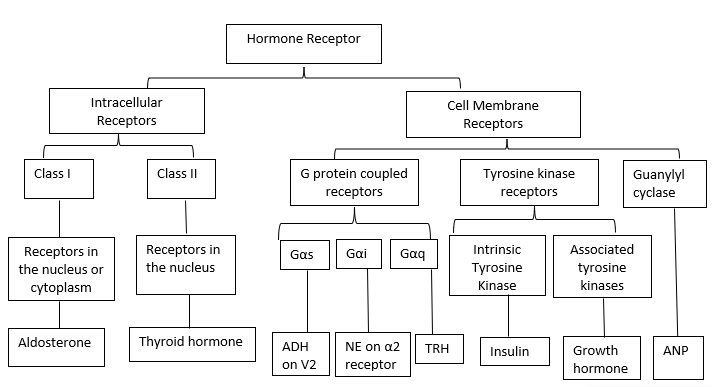
(B) Hormones act by binding to a specific protein called receptors that is located either on the cell membrane or inside the cell (cytoplasm or nucleus) of the target cells.
Classification of hormone receptors
2.
(A) The location of the receptors depends on the chemical structure of the hormone.
Receptors for peptide hormones and catecholamine’s are located in the cell membranes of target cells. Thyroid and steroid hormones cross the membrane and bind to receptors in the nucleus and cytoplasm. When hormones bind to their receptors, modifications in the receptor occur, resulting in an intracellular signaling system that triggers cellular responses such as increased protein synthesis, channel protein synthesis, cellular permeability alterations, and cell proliferation.
Hormones and their receptor’s location
Hormone
Class of Hormone
Receptor Location
Amine (epinephrine)
Water-soluble
Cell surface
Amine (thyroid hormone)
Lipid soluble
Intracellular
Peptide/protein
Water-soluble
Cell surface
Steroids and Vitamin D
Lipid soluble
Intracellular
3.
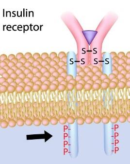
(D) Tyrosine Kinases is integral proteins within the plasma membrane. Example of tyrosine kinase receptors is insulin. The remaining second messengers are G protein coupled receptors (GPCRs).
When insulin for instance, binds to the receptor, it triggers 1. Dimerization of the receptor and signaling mechanism is triggered and the intrinsic tyrosine kinase is activated.
2. Tyrosine kinase activation enables phosphorylation of tyrosine residue within the receptor itself and intracellular tyrosine of target proteins.
3. Increase transportation of GLUT4 receptors through the cell to the cell membrane.
4. Increase the ability of the cells to uptake of glucose.
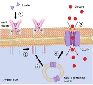
Activation of intrinsic tyrosine kinase
4.
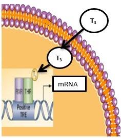
(B) thyroid hormones pass across the cell membrane and binds to its intracellular receptor found in the nucleus. Peptide hormone receptors (growth hormone, antidiuretic hormone and insulin) located in the cell membrane.
The activation steps of thyroid hormone receptors: 1. Thyroid hormones readily diffuse through the cell membrane.
2. Thyroid hormone interacts with the thyroid hormone receptor (THR), bound as a heterodimer with a retinoid acid X receptor (RXR) of the thyroid hormone response element (TRE) of the gene.
3. This result to either activation or inhibition of transcriptional proteins of that gene through changes in mRNA and therefore new protein formation in the cell.
Receptors of thyroid hormone are found inside the nucleus.
5.
(B) Hormones act by binding to a specific protein called receptors. This interaction will bring about changes in the receptor which result in intracellular signaling mechanism to initiate cellular responses that may include increase protein synthesis, formation of channel proteins, alteration in the cellular permeability, and cell growth (mitosis). A cellular response to hormone receptor binding does not result in cellular mutations.
6.
(A) Steroid’s hormones easily passing across the cell membrane and binds to intracellular receptor found in the cytoplasm or the nucleus.
The activation steps of steroid hormone receptors: 1. Steroid hormones diffuse easily into their target cells.
2. Steroid hormones bind and activate a specific intracellular receptor.
3. The hormone-receptor complex travels to the nucleus and dimerizes (not shown).
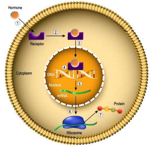
4. The hormone-receptor dimers binds to hormone response elements within the DNA.
5. This interaction prompts DNA transcription, to producing mRNA.
6. The new mRNA is translated into proteins.
7. Cellular effect by increasing cellular proteins and alteration of cellular functions.
For example, aldosterone induces the synthesis of sodium channels in the distal tubules and collecting ducts of the nephron.
Aldosterone, cortisol, progesterone, estrogen, and androgens are examples of steroid hormones that fall into this category.
Receptors of steroid hormones are found inside the cell.
7.
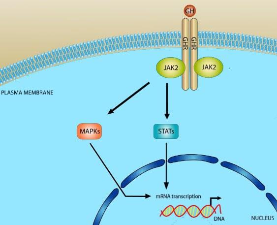
(B) Growth hormone receptors have single transmembrane spanning receptors that associated with tyrosine kinase such as Janus associated tyrosine kinase 2 (JAK2) The activation steps of growth hormone receptors: 1. Growth hormone binds to the receptor causes dimerization of the receptor 2. Migration of tyrosine kinase in associated proteins such as JAK2 towards the receptor.
3. A number of target proteins are activated including signal transducers and activators transcription (STAT), which causes formation of new mRNAs and synthesis of new proteins. JAK2 also phosphorylate other associated protein kinase family of mitogen-activated protein kinases (MAPK) which involves in cell growth and mitotic division.
Activation of tyrosine kinase-associated receptor
8.
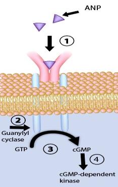
(A) Atrial natriuretic peptide (ANP) receptors activate guanylyl cyclase enzyme and it generates second messenger through cGMP.
Specification of peptide hormone receptors
G-protein
coupled Tyrosine
kinase Guanylyl
cyclase
receptors
receptors
associated receptors
Transmembran
Seven-pass
Single-pass
Single-pass
e
receptor receptor
receptors
receptors
types
Enzyme
Adenylyl
cyclase Tyrosine kinase
guanylyl cyclase
component
(Gs and Gi) and
phospholipase
C
(Gq)
Second
cAMP (Gs and Gi) No
second cGMP
messenger
and IP3 and DAG messenger
(Gq)
Example
9.
(D) Receptors for peptide hormones such as parathyroid hormone (PTH) are located in the cell membranes of target cells. Steroid hormones, thyroid hormones (thyroxine and triiodothyronine) and vitamin D (1, 25-dihydroxycholecalciferol) cross the membrane and upon their receptors located in the nucleus.
10.
(B) ADH act on its receptors V1a and V1b via second messenger and increases the levels of IP3 and DAG that result in vasoconstriction and increase release of ACTH
from the anterior pituitary gland respectively. ADH increases water reabsorption from the collecting tubule by binding to V2 receptors and increases the second messenger cAMP. The second messenger system is not present in tyrosine kinase.
Types, locations and actions of ADH (Vasopressin) receptors Types
of Receptors
Second
Actions
Receptors
Location
messenger
Vasopressin-
Vascular smooth Intracellular
Vasoconstriction
1
receptor/ muscle of blood calcium signaling (V1a)
vessels
Vasopressin-
Principal cells of Increase cAMP
Water reabsorption
2 receptor
the
kidneys
(V2)
collecting ducts
Vasopressin-
Anterior pituitary Intracellular
Stimulate release of
3 receptor/
gland
calcium signaling
ACTH
(V1b)
(Corticotroph)
HYPOTHALAMUS AND POSTERIOR PITUITARY HORMONES
1.
In terms of releasing hormones produced in the hypothalamus, which of the following statements is FALSE?
A.
They pass down hypothalamohypophysial system to reach the pituitary gland.
B.
May control the output of more than one pituitary hormone.
C.
Regulate the release of thyroid stimulating hormone (TSH).
D.
Regulate oxytocin storage and release.
2.
The two parts of the pituitary gland are the _____.
A.
infundibulum and neurohypophysis.
B.
neurohypophysis and adenohypophysis.
C.
adenohypophysis and pituitary stalk.
D.
infundibulum and hypothalamohypophysial portal system 3.
Which of the hormones are produced by the hypothalamus?
A.
Antidiuretic hormone (ADH).
B.
Follicle stimulating hormone (FSH).
C.
Thyroid stimulating hormone (TSH).
D.
adrenocorticotropic hormone (ACTH)
4.
Concerning the pituitary gland, which cell type is responsible for secreting both luteinizing hormone (LH) and follicle-stimulating hormone (FSH)?
A.
Lactotrophs cells.
B.
Thyrotrophs cells.
C.
Somatotrophs cells.
D.
Gonadotrophs cells.
5.
Which of the following is FALSE regarding antidiuretic hormone (ADH)?
A.
It is act on the arteriolar smooth muscle cells causing vasoconstriction.
B.
Increases the water permeability of the cells in the collecting tubule.
C.
Secretion is affected by changes in plasma osmolality.
D.
Secretion increases when plasma volume increase.
6.
All the following conditions trigger ADH secretion EXCEPT: A.
blood osmolality increases.
B.
blood pH increases.
C.
blood volume decreases.
D.
blood pressure decreases.
7.
The release of oxytocin occurs in response to_____.
A.
baby sucking during nursing.
B.
increased blood pressure.
C.
increased urine output.
D.
a hypothalamic-releasing hormone.
8.
All the following are anterior pituitary hormones EXCEPT: A.
Growth hormone (GH).
B.
Adrenocorticotrophic hormone (ACTH).
C.
Prolactin inhibitory hormone (PIH).
D.
Follicle stimulating hormone (FSH).
Which hormone causes the contraction of smooth muscle cells in the uterus, leading to the initiation of labor and childbirth?
A.
Adrenocorticotropic hormone (ACTH)
B.
Follicle stimulating hormone (FSH)
C.
Luteinizing hormone (LH)
D.
Oxytocin
10.
The hypothalamus is functionally and anatomically connected to the posterior pituitary lobe by a bridge of _____.
A.
blood vessels
B.
bone
C.
nerve axons
D.
cartilage
Answers:
1.
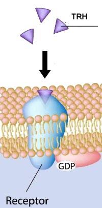
(D) The hypothalamus produces oxytocin, but the posterior pituitary gland stores and releases it. The hypothalamic-hypophyseal portal system is a vascular connection between the hypothalamus and anterior pituitary gland. Releasing hormone from the hypothalamus (such as TRH, CRH, GnRH, GHRH) travel through the hypothalamic-hypophyseal portal system where they act upon specific membrane receptors within the anterior pituitary gland and stimulate synthesis and secretion of anterior pituitary hormones. Thyroid releasing hormone (TRH) stimulate the release of Thyrotropin (also known as thyroid stimulating hormone).
Hypothalamic releasing and inhibitory hormones that pass to the anterior pituitary from the median eminence via the hypothalamo-hypophyseal portal vessels Hypothalamic hormones and their actions
Hypothalamic hormones
Actions of hypothalamic
hormones on anterior pituitary
Thyrotropin releasing hormone: (TRH)
Stimulate release of TSH
stimulation of prolactin secretion
Gonadotropin
releasing
hormone Stimulate release of LH and FSH
(GnRH)
Corticotropin releasing hormone (CRH)
Stimulate release of ACTH
Growth
hormone-releasing
hormone Stimulate secretion of growth
(GHRH)
hormone (GH)
Growth
hormone-inhibitory
hormone Inhibit the release of GH
(GHIH) also known as Somatostatin
Prolactin-inhibitory factor (PIF) also Inhibit release of Prolactin known as dopamine
2.
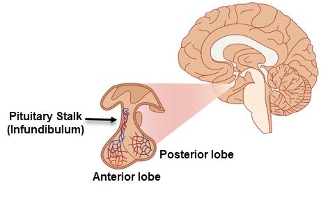
(B) The posterior pituitary gland is called neurohypophysis, while the anterior pituitary gland is called adenohypophysis. The pituitary stalk also known as the infundibulum, connects the base of the hypothalamus with the pituitary gland.
Pituitary gland anatomical parts
3.
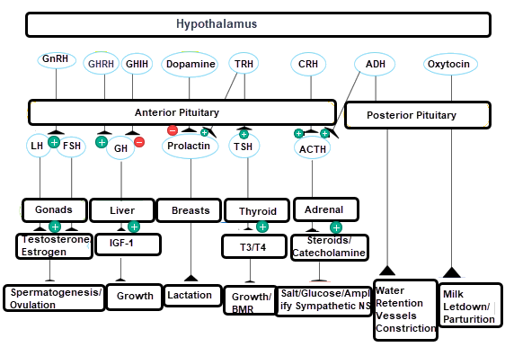
(A) The hypothalamus produces antidiuretic hormone (ADH). The anterior pituitary gland makes FSH, TSH, and ACTH.
The role of hypothalamus as a master of endocrine glands 4.
(D) Gonadotrophs are basophilic cells of the anterior pituitary gland specialized to secrete gonadotropins in response to hypothalamic gonadotropin-releasing hormone (GnRH). Lactotrophs are acidophilic cells in the anterior pituitary. They produce and release prolactin (PRL) in response to thyrotropin-releasing hormone. The hormone dopamine causes inhibition of PRL secretion. Thyrotrophs are the cells in the anterior pituitary that release thyroid-stimulating hormone (TSH) in response to thyrotropin releasing hormone (TRH) produced by the hypothalamus. Somatotrophs are the cells in the anterior pituitary that releases growth hormone (also called somatotropin).
Hypothalamic hormones and the anterior pituitary gland's target cells
Hypothalamic hormones
Target cells in the
Actions of hypothalamic
anterior pituitary
hormones on anterior
pituitary
1.
Thyrotropin
releasing
hormone: Thyrotrophs
Stimulate release of TSH
(TRH)
Lactotrophs
stimulation
of
prolactin
secretion
2.
Gonadotropin releasing hormone Gonadotrophs Stimulate release of LH and
(GnRH)
FSH
3.
Corticotropin
releasing
hormone Corticotrophs
Stimulate release of ACTH
(CRH)
4.
Vasopressin (ADH)
Corticotrophs
Stimulate release of ACTH
5.
Growth hormone-releasing hormone Somatotrophs Stimulate secretion of growth
(GHRH)
hormone (GH)
6.
Somatostatin also known as Growth Somatotrophs Inhibit the release of GH
hormone-inhibitory hormone (GHIH)
7.
Dopamine also known as Prolactin-
Lactotrophs
Inhibit release of Prolactin
inhibitory factor (PIF)
(D) Antidiuretic hormone (ADH) is a polypeptide hormone, synthesized from the hypothalamus. Stored in secretory granules and secreted from the posterior pituitary gland. Secretion of ADH increases when plasma volume and blood pressure decrease. High plasma osmolality also increases release of ADH. ADH acts on the principal cells of the nephron cortical collecting duct and stimulate water reabsorption. ADH acts on the vascular smooth muscle resulting in vasoconstriction and increasing blood pressure. ADH act along with corticotropin releasing hormone to stimulate the release of ACTH from the anterior pituitary.
6.
(B) ADH secretion is unaffected by blood pH. ADH secretion is stimulated by changes in plasma osmolality and blood volume.
Regulation of ADH secretion
Factors stimulate ADH secretion
Factors inhibit ADH secretion
1. Hyperosmolarity
Hypo-osmolarity
2. Low blood volume
High blood volume
3. Low blood pressure
High blood pressure
7.
(A) Oxytocin plays a role in milk ejection. The milk is stored in mammary alveoli and small milk ducts. The major action of oxytocin is to cause milk letdown. Sucking of the baby stimulate sensory neuron from the nipple to relay signal to the hypothalamus which activate paraventricular and supraoptic nucleus that causes action potential to travels along the neural tract to the posterior pituitary, resulting release of oxytocin. Oxytocin acts on breast ducts cause smooth muscle contraction and milk letdown.
Schematic illustration representing the mechanism of oxytocin secretion Baby sucking
Sensory nerve
Hypothalamus
Action potential
Posterior pituitary
Stimulate release
Oxytocin
Acts on smooth muscle in milk ducts
causing milk ejection
(C) Prolactin inhibitory hormone (PIH) is a hypothalamic hormone that inhibit the secretion of prolactin from the anterior pituitary gland. The remaining hormones are pituitary hormone.
Posterior lobe (Neurohypophysis): Neural tissue that receives, stores, and releases hormones from the hypothalamus such as antidiuretic hormone and oxytocin.
Anterior lobe (Adenohypophysis): Made up of a variety of cell types which secrete hormones.
Anterior and posterior pituitary hormones
Anterior pituitary (adenohypophysis)
Posterior pituitary (neurohypophysis)
Luteinizing hormone (LH)
Oxytocin
Follicle Stimulating Hormone (FSH)
Vasopressin
(Antidiuretic
Hormone;
Thyroid Stimulating Hormone (TSH)
ADH)
Growth Hormone (GH)
Adrenocorticotropic Hormone (ACTH)
Prolactin (Prl)
9.
(D) During labor, the hormone oxytocin is released, which triggers uterine smooth muscle contractions. Oxytocin can be used clinically to assist the birth process.
ACTH, FSH and LH all are anterior pituitary hormones. ACTH stimulate the release steroid hormones. FSH and LH control the release of the sex hormones testosterone and estrogen in males and females, respectively.
Schematic diagram depicting the mechanism of oxytocin action during childbirth Stretch of cervix
Sensory nerve
Hypothalamus
Action potential
Posterior pituitary
Stimulate release
Oxytocin
Stimulate contraction of
uterine smooth muscle
10.
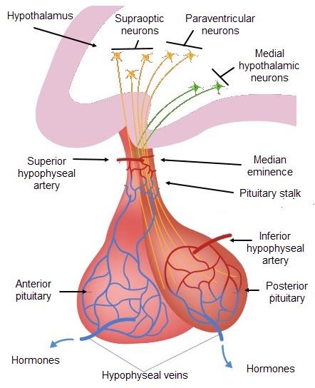
(C) A neuronal connection exists between the hypothalamus and the posterior pituitary. Neuron cell bodies in the hypothalamus’s supraoptic and paraventricular nuclei extend its axonal extensions to the posterior pituitary.
Neural relationship between the hypothalamus and the posterior pituitary. Neurons cell bodies in the supraoptic and paraventricular nuclei of the hypothalamus have their axonal extension to the posterior pituitary. Vascular relationship between the hypothalamus and the anterior pituitary. Several neurons of the medial hypothalamic nuclei produce pituitary releasing hormones that project to the median eminence, where they pass via portal vein to influence secretion of anterior pituitary hormones.
Anterior pituitary – Growth hormone and prolactin 1.
Regarding growth hormone (GH), all the following statements are correct EXCEPT:
A.
Act indirectly by stimulating IGF-1
B.
It secreted at a constant rate throughout the day.
C.
Increase lipolysis
D.
It has diabetogenic effect
2.
Failure of the pituitary to stop producing growth hormone after body growth is completed results in _______.
A.
Gigantism
B.
Cushing’s syndrome
C.
Dwarfism
D.
Acromegaly
3.
The polypeptide growth factors secreted by the liver is _____.
A.
Growth hormone releasing hormone (GHRH)
B.
Somatostatin
C.
Growth hormone
D.
Somatomedins
4.
All of the following statements about growth hormone regulation are true EXCEPT: A.
Is stimulated by somatostatin released from the hypothalamus.
B.
It rises when the blood glucose level declines.
C.
Increases the size of viscera.
D.
Stimulates the formation of somatomedins in the liver.
5.
Which of the following pituitary hormones has a chemical structure most similar to that of growth hormone?
A.
Follicle stimulating hormone
B.
Adrenocorticotropic hormone
C.
Thyroid-stimulating hormone
D.
Prolactin
6.
Which of the following circumstances would growth hormone secretion most likely suppress?
A.
Low somatostatin concentration.
B.
High somatomedins (IGF – 1) concentration
C.
High GHRH concentration.
D.
Low glucose concentration.
7.
The metabolic effect of growth hormone includes all of the following EXCEPT: A.
Protein synthesis
B.
Glucose utilization
C.
Lipolysis
D.
Gluconeogenesis
Which of the following is TRUE regarding prolactin?
A.
Has a similar chemical structure to cortisol.
B.
Release is stimulated by dopamine.
C.
Secretion is stimulated by suckling of the breast.
D.
Causes pre-formed milk to be ejected by the breast during suckling.
9.
Which of the following statements about lactation is FALSE?
A.
Milk formation is stimulated by estrogen and progesterone.
B.
Milk formation can be depressed by hypothalamic activity.
C.
Maintenance of lactation depends on suckling.
D.
Lactation ceases if the anterior pituitary gland is destroyed.
10.
Which of the following describe excessive lactation in women due to inappropriate prolactin secretion?
A.
Galactorrhea
B.
Amenorrhea
C.
Oligomenorrhea
D.
Endometriosis
Answers:
1.
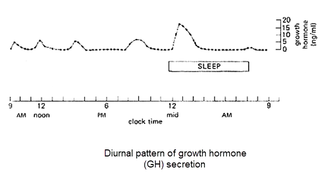
(B) GH secretion has a diurnal pattern, with levels increasing during the early night and during sleep. Growth hormone (GH) is secreted by the anterior pituitary gland in a pulsatile fashion under the regulation of two hypothalamic hormones: GH-releasing hormone (GHRH) stimulates GH synthesis and secretion while somatostatin inhibits GH release.
Hormone can be secreted in response to:
Hormonal Stimuli
Response
Humoral
stimuli:
release
of Secretion of insulin in response to high hormones in response to nutrient in glucose level.
the blood.
Secretion
of
Parathyroid
hormone
in
response to low calcium level.
Neural
stimuli:
Secretion
of Secretion Epinephrine and norepinephrine hormone in response to neural from the adrenal medulla increased by high activity.
sympathetic nervous system activity.
Hormonal secretion: release of Secretion of thyroid hormones (T3 and T4) in hormones
in
response
to response to TSH release.
hormones
produced
by
other
endocrine organs.
Diurnal
secretion:
Hormone Melatonin
secretion
increased
during
secretion fluctuates according to darkness and inhibited during daylight.
exposure to light.
Growth hormone secretion increase during
the night and decreased in the daytime.
2.
(D) Acromegaly is the excess secretion of growth hormone that develops during adulthood after epiphyseal plates close. Enlargement of extremities (hands and feet) and face, thickening of soft tissue. Gigantism: excess secretion occurs before adolescence before epiphyseal plates close. Dwarfism: deficiency of anterior pituitary secretion during childhood. Cushing's syndrome is a disorder that occurs due to hypersecretion of glucocorticoid.
3.
(D) Somatomedins (insulin like growth factor 1) are a group of peptide hormone.
Somatomedin’s production is stimulated by the action of growth hormone on liver
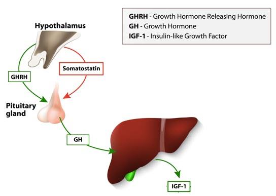
cells (hepatocytes). GHRH is a releasing hormone of growth hormone. Somatostatin also known as growth hormone-inhibiting hormone (GHIH) inhibits the secretion of growth hormone
Somatomedins (IGF-1) secretion from the liver cells by the action of growth hormone.
4.
(A) Growth hormone is secreted in response to hypothalamic growth hormone releasing hormone (GHRH) and inhibited by Somatostatin (growth hormone inhibitory hormone, GHIH) produced by the hypothalamus.
Regulation of growth hormone secretion
Growth hormone stimulation
Growth hormone inhibition
Growth hormone releasing hormone
Somatostatin (GHIH)
(GHRH)
Decrease blood glucose
Increase blood glucose (Hyperglycemia)
(Hypoglycemia)
Decrease blood free fatty acids
Rising blood free fatty acids
Exercise
Somatomedins (IGF)
Deep sleep
Inhibited during day time
Stress
5.
(D) Both growth hormone and prolactin are large polypeptides. Growth hormone has 191 amino acids with two disulfide bonds. While prolactin has 199 amino acids with three disulfide bonds.
6.
(B) High somatomedin levels reduce GH secretion due to its negative feedback mechanism on both the hypothalamus and the pituitary gland. Low somatostatin (GHIH) increases growth hormone secretion. High GHRH and low blood glucose concentration (hypoglycemia) are stimuli that increase growth hormone secretion.
Low somatomedin levels increase GH secretion.
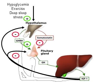
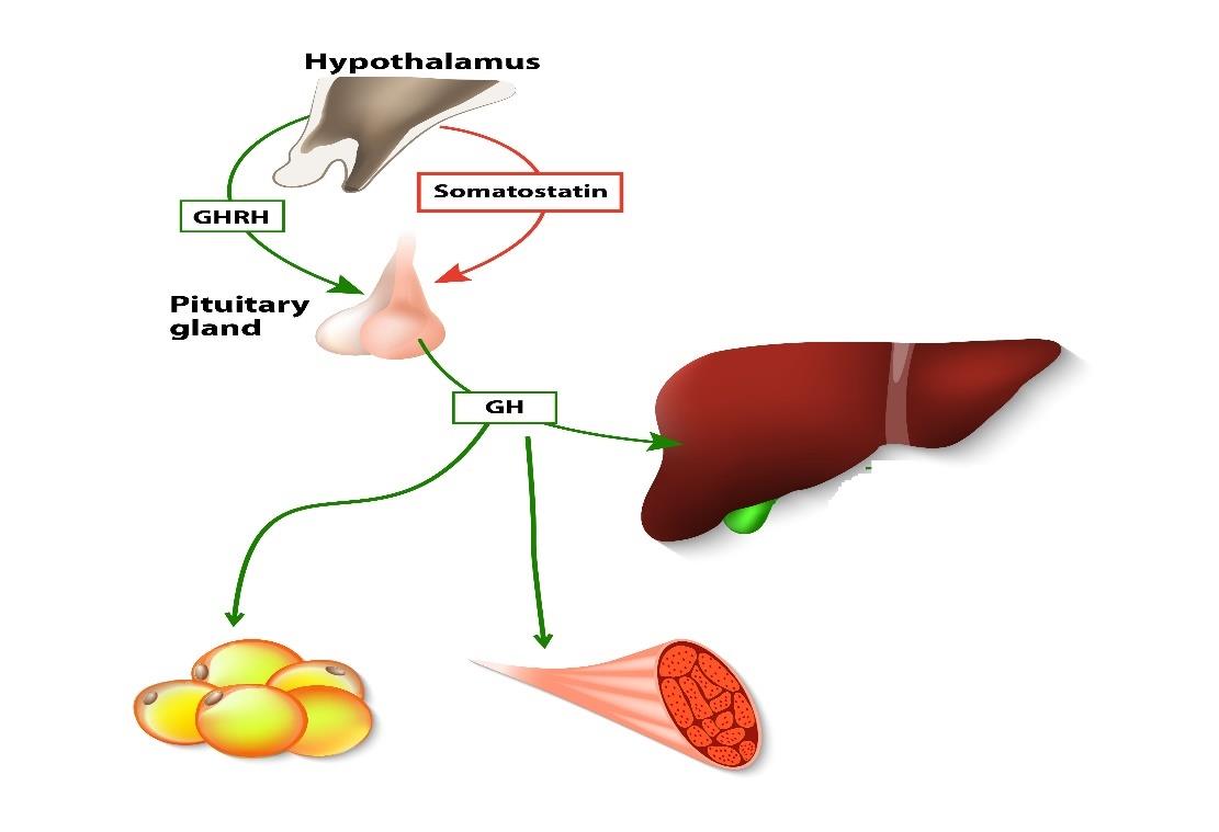
Regulation of growth hormone (GH) secretion 7.
(B) GH have an anabolic and catabolic actions. In terms of proteins, GH increases protein synthesis (anabolic effect) and reduces the breakdown of cell proteins by decreasing catabolism of protein. GH Causes release of fatty acids from adipose tissue and then increasing the concentration of fatty acids. In terms of carbohydrates, GH inhibits glucose uptake by cells and increases gluconeogenesis, causing blood glucose levels to rise (Catabolic).
Metabolic actions of growth hormone (GH)
8.
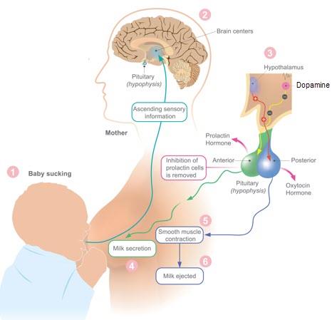
(C) Baby sucking stimulates prolactin secretion from the anterior pituitary gland by inhibiting dopamine release from the hypothalamus, as dopamine inhibits prolactin secretion. Prolactin is a polypeptide hormone, while cortisol is a steroid hormone, hence their structures are different. The production of oxytocin causes milk ejection.
The steps involved in releasing and the action of prolactin 1. Baby sucking.
2. Inhibition of dopamine release from the hypothalamus.
3. Release of prolactin from the anterior pituitary.
4. Initiates lactation.
The steps involved in releasing and the action of oxytocin 1. Baby sucking
2. Ascending sensory information to the hypothalamus.
3. Synthesis and release of oxytocin by the hypothalamic neuron (Paraventricular and supra-optic nuclei).
4. Oxytocin acts on the smooth muscle in the mammary duct to eject milk.
The role of prolactin and oxytocin and their effect in milk secretion and milk ejection 9.
(A) Milk formation is stimulated by prolactin. Prolactin inhibits GnRH secretion and thus reduce the plasma levels of estrogen and progesterone.
Prolactin is regulated by the release of prolactin-inhibiting hormone (dopamine) from the hypothalamus. Factors that inhibit dopamine such as baby sucking stimulate the release of prolactin.
The major stimulus to prolactin secretion is suckling which initiates and maintains lactation after delivery.
Milk formation ceases due to loss of prolactin.
10.
(A) Galactorrhea is the term used to describe excessive lactation in women caused by inappropriate secretion of prolactin. The most common cause of hyperprolactinemia is a prolactin secreting pituitary tumor. Women present with galactorrhea associated with a range of menstrual disturbances including oligomenorrhea (infrequent menstrual periods) and amenorrhea (absence of menstruation).
1.
The secretion of adrenocorticotrophic hormone (ACTH) is DECREASED in which of the following conditions?
A.
When the level of corticotropin releasing hormone (CRH) rise.
B.
When the amount of cortisol in the blood increases.
C.
When you are about to get up in the morning.
D.
Following severe traumatic event.
2.
Which of the following clinical features is related with hyposecretion of adrenal cortex hormones?
A.
Hypertension.
B.
Hyperglycemia.
C.
Hyperkalemia.
D.
Hypernatremia.
3.
Injections of cortisol trigger a rise in all the following EXCEPT: A.
Interleukin-1.
B.
Bone resorption.
C.
Blood pressure.
D.
Catabolic activity in muscle.
4.
Regarding cortisol, choose the FALSE statement.
A.
Cortisol is bound to plasma protein in the blood.
B.
Cortisol is a peptide hormone.
C.
Cortisol blood levels peak in the morning.
D.
Cortisol Inhibits release of ACTH from the anterior pituitary gland.
5.
The main secretory products of the adrenal medulla are _____.
A.
glucocorticoids.
B.
mineralocorticoids.
C.
androgens.
D.
catecholamines.
6.
All the following conditions increase aldosterone secretion EXCEPT: A.
ACTH concentration.
B.
Osmolality.
C.
Potassium concentration.
D.
Renin concentration.
7.
Adrenaline secretion from the adrenal medulla ______.
A.
decreases blood glucose level.
B.
decreases free fatty acid level.
C.
increases blood flow to skeletal muscle.
D.
increases renal blood flow.
8.
All are features of adrenal cortex hormones EXCEPT: A.
Cholesterol is the main source of steroid hormone synthesis.
B.
Are mostly bound to plasma proteins.
C.
Over-secretion causes phaeochromocytoma.
D.
Include sex hormones.
The plasma level of adrenocorticotrophic hormone (ACTH) A.
Is normally highest around midnight.
B.
Is regulated mainly by the blood cortisol level.
C.
Shows circadian fluctuations.
D.
Is raised in patients on long-term high dosage glucocorticoids.
10.
The primary stimulus for release of adrenal medullary hormones is _____.
A.
the kidneys.
B.
the sympathetic nervous system.
C.
the anterior pituitary.
D.
the parasympathetic nervous system.
Answers:
1.
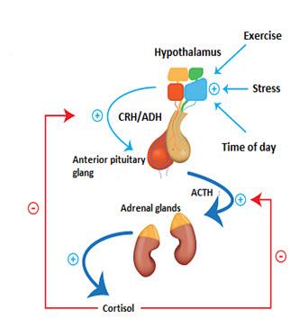
(B) ACTH acts on the cortex of the adrenal glands to stimulate the synthesis of cortisol. Negative feedback by cortisol makes the cells that produce ACTH
(corticotrophs) less responsive to CRH. Corticotrophin releasing hormone (CRH) stimulate the secretion of Adrenocorticotrophic hormone (ACTH). ACTH has a pulsatile and diurnal secretory pattern that drives a parallel pattern of cortisol secretion. The lowest secretion is during the evening hours and after falling asleep, and the highest secretory rates occur just before awakening in the morning. Stress, trauma and emotions stimulate ACTH secretion.
2.
(C) Hypo-secretion of glucocorticoids and mineralocorticoids results in increased blood K+ (hyperkalemia), decreased Na+ (hyponatremia), low blood volume (hypotension), decreased blood glucose levels (hypoglycemia).
3.
(A) Cortisol inhibits the production of interleukin-1 (IL-1) from the macrophages and inhibits the production of interleukin-2 (IL-2) and the proliferation of T lymphocytes.
Cortisol inhibits the release of histamine and serotonin from mast cells and platelets.
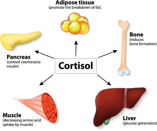
Cortisol increases protein catabolism in muscle and decreases new protein synthesis, thereby providing additional amino acids to the liver for gluconeogenesis.
Cortisol maintains vascular responsiveness to catecholamines and thus elevate blood pressure and increase bone resorption and inhibit bone formation.
4.
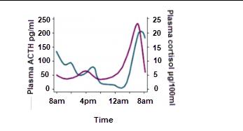
(B) Cortisol is a steroid hormone not a peptide hormone. Steroid hormones are mostly bound to plasma proteins. The lowest secretion is during the evening hours and after falling asleep, and the highest secretory rates occur just before awakening
in the morning Cortisol Inhibits release of ACTH from the anterior pituitary gland by negative feedback mechanism.
5.
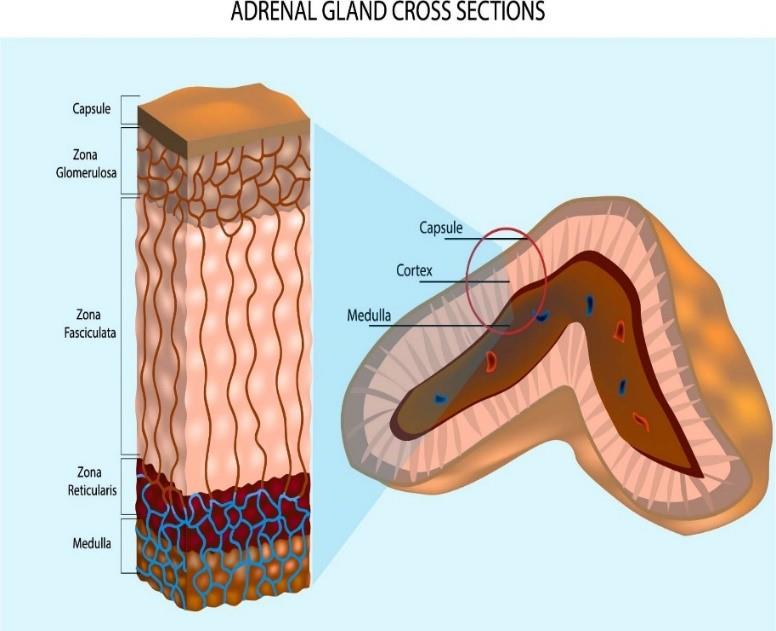
(D) Adrenal medulla releases epinephrine and norepinephrine (catecholamines) into blood. The adrenal cortex secretes three classes of steroid hormones: glucocorticoids, mineralocorticoids, and androgens. Each layer of the adrenal cortex synthesizes one of the three types of hormones. The innermost zone of the cortex called the zona reticularis, secretes mainly adrenal androgens. The middle called the zona fasciculata, secretes glucocorticoids. The outermost zone, called the zona glomerulosa, secretes mineralocorticoids.
6.
(B) Change in osmolarity stimulate ADH secretion. The action of ACTH is on glucocorticoid secreting cells; it has some action on mineralocorticoid secreting cells.
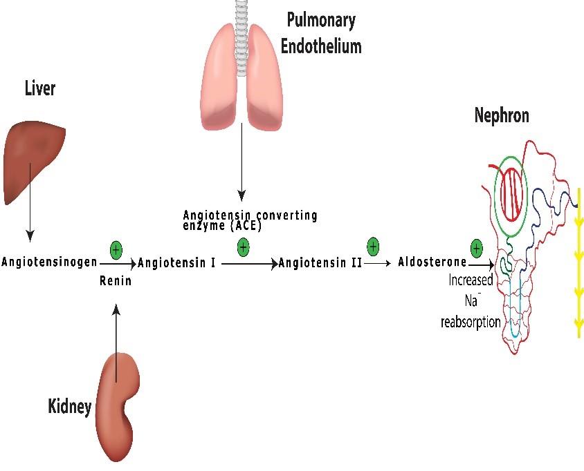
K+ has a direct stimulatory effect on the adrenal cortex. Increase renin leads to formation of angiotensin II which stimulates the cortex to release aldosterone.
7.
(C) By its effect on β2 receptors in the smooth muscle of skeletal muscle arterioles.
Adrenaline increases blood glucose and fatty acid by promoting glycogenolysis,
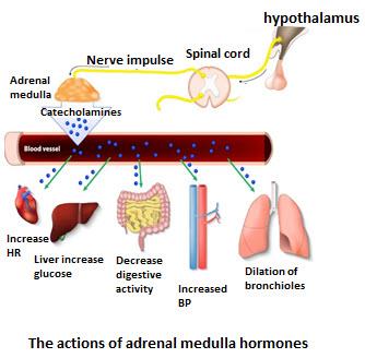
gluconeogenesis and lipolysis. Adrenaline reduces renal blood flow by stimulating α1
receptors result in vasoconstriction.
8.
(C) Phaeochromocytoma is over secretion of catecholamines due to tumor within the adrenal medulla. Cholesterol is the main source of steroid hormone synthesis. All of the steroids are chemical modifications of a cholesterol. Steroid hormones bind to plasma proteins. Zona reticularis produces gonadocorticoid such as androgens.
9.
(C) ACTH shows circadian fluctuations. The lowest secretion is during the evening hours and after falling asleep, and the highest secretory rates occur just before awakening in the morning. Due to negative feedback mechanism, ACTH levels drop when taking high doses of glucocorticoids for a long time.
Diurnal pattern of adrenocorticotropic (ACTH) hormone and cortisol secretion
10.
(B) Adrenal medulla hormones secretions increased when stimulated by increased sympathetic activity. Output is increased by stress, fear, cold, hypoglycemia, blood loss.
1.
Thyroid hormones, when secreted in large amounts, they can cause a rise in all of the following EXCEPT:
A.
Basal metabolic rate.
B.
Total peripheral resistance.
C.
Heart rate and contraction strength.
D.
Gastrointestinal motility and absorption.
2.
Which of the following does thyroid-stimulating hormone (TSH) secretion decrease?
A.
In primary hyperthyroidism seen in Graves' disease.
B.
In children with congenital hypothyroidism.
C.
In hypothyroid condition seen in Hashimoto's thyroiditis.
D.
Surgical removal of thyroid gland.
3.
All of the following statements about the thyroid gland is true EXCEPT: A.
It is located anterior to the trachea and inferior to the larynx.
B.
The parathyroid glands are located at the back of it.
C.
It produces thyroxine, triiodothyronine and calcitonin.
D.
It has relatively low blood perfusion.
4.
Thyroid hormone production is regulated by __________.
A.
corticotropin-releasing hormone (CRH) from the hypothalamus B.
thyroid stimulating hormone (TSH) from the anterior pituitary C.
thyroxine (T4) from the anterior pituitary
D.
thyroglobulin (Tg) from the thyroid’s parafollicular cells 5.
Iodide enters thyroid follicular cells by which of the following mechanisms?
A.
simple diffusion
B.
facilitated diffusion
C.
active transport
D.
osmosis
6.
In thyroid gland follicles, a glycoprotein stored in colloid is _____.
A.
thyroglobulin (Tg)
B.
thyroxine (T4)
C.
triiodothyronine (T3)
D.
thyroid binding globulin (TBG)
7.
Which of the following findings is most likely to be seen in a hypothyroid myxedema patient?
A.
Heat intolerance.
B.
Increased appetite.
C.
Reduced cholesterol.
D.
Weight gain.
8.
Which of the following events concerning thyroid hormone synthesis occurs last?
A.
The lysosome fuses with the endocytosed colloid.
B.
Iodine attaches to tyrosine residues in thyroglobulin.
C.
Iodides (I–) are actively taken into the cell.
D.
Thyroglobulin is synthesized and discharged into the colloid.
Select the TRUE statements with regard to thyroid physiology A.
T3 has greater secretion rate from the thyroid than T4
B.
T3 and T4 bind and act at the same cell membrane receptor C.
T3 and T4 are synthesized from tyrosine held in thyroglobulin D.
T4 is more active than T3
10.
Regarding thyroxine, which of the following is INCORRECT?
A.
Increases number of β adrenergic receptors on the heart B.
Thyroid hormones decrease carbohydrate absorption by the stomach and small bowel
C.
Hyaluronic acid accumulates in the skin if there is a deficiency of thyroxine D.
Large doses can increase body temperature
Answers:
1.
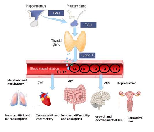
(B) Thyroid hormone affects the cardiovascular system by lowering total peripheral resistance, causing vasodilation, and increasing heart rate and force of contraction, resulting in an increase in cardiac output. Thyroid hormone raises the amount and activity of mitochondria, which raises the basal metabolic rate. Thyroid hormone increases the motility, absorption, and secretion of the gastrointestinal tract. Thyroid hormones are involved in the development of the fetal central nervous system as well as skeletal growth in children.
Actions of thyroid hormones
Body System
Actions
Respiratory system
Increases respiratory rate
Cardiovascular system
Increases heart rate and reduce total
peripheral resistance
Gastrointestinal system
Increases motility and absorption
Central nervous system
Growth and development
Reproductive system
Enhance reproductive functions
Metabolism
Increases basal metabolic rate (BMR)
2.
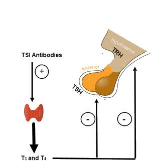
(A) Thyroid stimulating hormone (TSH) levels fall in hyperthyroidism situations such Graves' disease as a result of pituitary suppression by circulating thyroxine. In Grave’s disease, thyroid stimulating antibodies (TSI) are directed against thyroid stimulating hormone (TSH) receptors located in the basolateral membrane of thyroid cells. TSH receptors are stimulated by autoantibodies, resulting in increased T3 and T4 synthesis.
High levels of TSH are seen in hypothyroid diseases such as Hashimoto’s thyroiditis, surgical removal of thyroid gland (thyroidectomy) and congenital hypothyroidism due to congenital absence or underdevelopment of the thyroid gland during fetal development.
Increased levels of thyroid hormones, signal hypothalamus and anterior pituitary to inhibit secreting of TRH and TSH. TSI (Thyroid-Stimulating Immunoglobulin).
3.
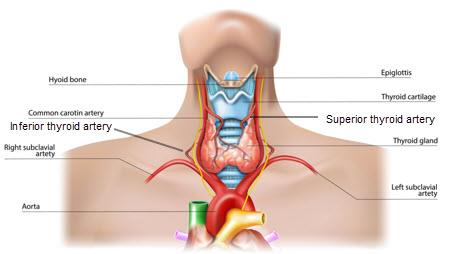
(D) The blood flow rate of the thyroid gland is one of the highest in the body. The thyroid gland receives blood supply from two major arteries: the superior thyroid artery and the inferior thyroid artery. An additional artery, the thyroid ima artery, is found in approximately 10% of the population. Thyroid gland is located anterior to the trachea and below to the larynx. Thyroid gland produces three hormones namely thyroxine, triiodothyronine and calcitonin. There are usually four parathyroid glands located behind the thyroid gland.
Thyroid gland location and blood supply
4.
(B) Thyroid stimulating hormone (TSH) is produced by Thyrotrophs, which are cells found inside the anterior pituitary gland. Corticotropin-releasing hormone (CRH) is a hormone secreted by the hypothalamus that regulates the secretion of adrenal cortex hormones. Thyroglobulin is a protein that is produced by the thyroid gland's follicular cells and is required for the production of two thyroid hormones: triiodothyronine (T3) and thyroxine (T4).
Regulation of thyroid hormone secretion
5.
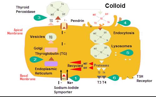
(C) An active transport mechanism transports iodide into thyroid follicular cells (thyrocytes) along with sodium against its electrochemical gradient via a sodium/iodide symporter (NIS). Iodide is transported out of the cell into the colloid via the iodide transporter called pendrin.
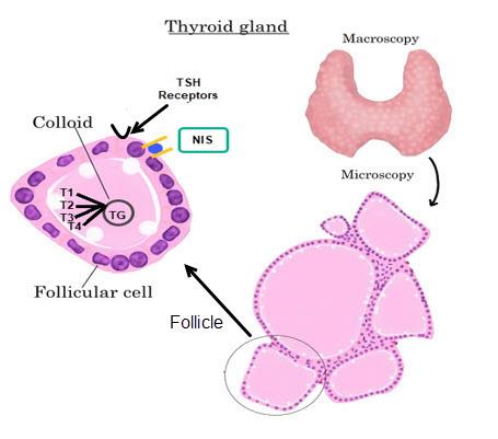
Histological features of thyroid gland
6.
(A) Thyroglobulin (Tg) acts as a substrate for the synthesis of the thyroid hormones thyroxine (T4) and triiodothyronine (T3), as well as the storage of the inactive forms of thyroid hormone and iodine (T1 and T2) within the follicular lumen of a thyroid follicle. T3 and T4 are the two main thyroid hormones. Thyroid binding globulin (TBG) is a plasma protein produced in the liver and binds thyroid hormones (T3 and T4) and carries them in the circulation.
7.
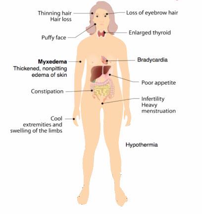
(D) Hypothyroidism is associated with weight gain despite poor appetite.
Hyperthyroidism is characterized by heat intolerance, increased appetite, and lower cholesterol levels.
Signs and symptoms of hypothyroidism
Body System
Symptoms
Integumentary system
skin thickening without pitting edema (myxedema).
Dry and inelastic skin develops.
Hair patterns and eyebrows change.
Cardiovascular System
Decreased heart rate (Bradycardia)
Decreased cardiac output
Fluid retention
Gastrointestinal System
Weight gain
Decreased gut motility and constipation
Central Nervous System
Decreased mental stability and forgetfulness
Sensitivity to the cold
Depression
Myxedema coma
Reproductive system
Menorrhagia
Decreased libido
8.
(A) The following are the steps involved in the synthesis of thyroid hormones: 1. Iodides (I–) are actively taken via a sodium/iodide symporter (NIS) into the cell and released into the colloid. 2. Thyroglobulin is synthesized and discharged into the colloid. 3. Iodides attaches to tyrosine within the thyroglobulin which is mediated by thyroperoxidase enzymes, forming T1 (monoiodotyrosine, or MIT), and T2
(diiodotyrosine, or DIT). 4. Iodinated tyrosine residues link together to form T3 and T4.
5. Colloid is then endocytosed and fused with a lysosome, where T3 and T4 are cleaved and diffuse into the bloodstream. 6. The release of T3 and T4 to the circulation. Thyroglobulin that remains is recycled and used to make additional colloid in the thyrocytes.
Steps of synthesis of thyroid hormones
9.
(C) T3 and T4 are synthesized from tyrosine within the thyroglobulin molecule in the colloid of the thyroid follicles. Both T3 and T4 act on the nuclear receptors and not cell membrane receptors. The secretion rate and plasma concentration are higher in T4 than T3. T3 is more metabolically active hormone than T4.
(B) Thyroxine increases carbohydrate absorption by the stomach and gut motility.
Increased dermal glycoaminoglycan and hyaluronic acid content traps water and makes the skin coarse and thickened (myxedema) are the clinical manifestation seen in patient with hypothyroidism. Large doses of thyroxine increase the basal metabolic rate and heat production and thus body temperature.
1.
All of the following are the physiological actions of testosterone EXCEPT: A.
Depresses pituitary secretion of LH.
B.
Causes the epiphyses of long bones to unite.
C.
Skeletal muscle hypertrophy.
D.
Stimulates growth of scalp hair.
2.
For normal development and fertility of spermatozoa there must be A.
Secretion of testosterone.
B.
Secretion of luteinizing hormone.
C.
Secretion of follicle-stimulating hormone.
D.
A testicular temperature of 370C.
3.
All of the statements about testosterone secretion from the testis are correct EXCEPT:
A.
Increases at puberty because LH levels increase.
B.
Peaks in the early evening.
C.
Has a negative feedback effect on LH secretion by the anterior pituitary gland.
D.
Is responsible for the growth of facial hair.
4.
In a female menstrual cycle, ovulation produces all of the following hormonal changes. EXCEPT:
A.
Estrogen production is very high.
B.
Formation of corpus luteum.
C.
high production of follicle-stimulating hormone.
D.
high production of luteinizing hormone.
5.
In the normal menstrual cycle, which of the following is FALSE?
A.
The proliferative phase depends on estrogen secretion.
B.
Cervical mucus becomes more fluid around the time of ovulation.
C.
Ovulation is followed by a surge in blood luteinizing hormone level.
D.
Basal body temperature is higher after ovulation.
6.
Seven days after ovulation, pituitary secretion of luteinizing hormone (LH) decreases rapidly. What is the cause of this decrease in secretion?
A.
The anterior pituitary gland becomes unresponsive to the stimulatory effect of gonadotropin releasing hormone (GnRH).
B.
Estrogen from the developing follicles exerts a feedback inhibition on the hypothalamus.
C.
The rise in body temperature inhibits hypothalamic release of GnRH.
D.
Secretion of estrogen and progesterone by the corpus luteum suppresses hypothalamic secretion of GnRH and pituitary secretion of LH.
7.
Select the TRUE statement regarding the human chorionic gonadotrophic hormone (HCG):
A.
It is a steroid hormone.
B.
Can be detected in the urine as an early sign of pregnancy.
C.
Blood level rises steadily throughout pregnancy.
D.
It acts directly on the uterus to maintain the endometrium.
Secondary amenorrhea (disappearance of previously established menstruation) occurs in the following conditions EXCEPT:
A.
A female body fat content of approximately 25%.
B.
Severe weight loss.
C.
Continuous administration of gonadotropin-releasing hormone (GnRH).
D.
A strenuous daily training schedule.
9.
The followings are the symptoms of menopause EXCEPT: A.
Vaginal dryness.
B.
Hot flashes and night sweats.
C.
Libido (sex drive) may increase.
D.
Osteoporosis.
10.
Which of the following steroid hormones does the fetus provide for the placenta to produce estrogen?
A.
Androstenedione.
B.
Dehydroepiandrosterone sulfate (DHEA-S).
C.
Dihydrotestosterone.
D.
Testosterone.
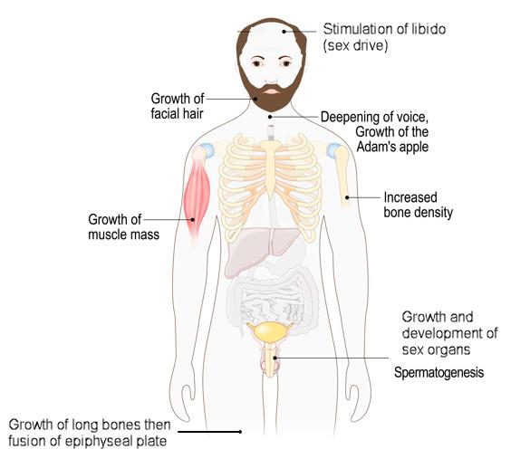
Answers:
1.
(D) During puberty, the action of testosterone often results in a temporal recession of the hair line. Testosterone is an anabolic hormone that promote an increase in the muscle mass. Testosterone contribute to the growth of long bones and causing the fusion of epiphyseal plates and thus ceasing the growth of long bones.
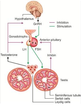
The hypothalamo-pituitary-testicular axis is regulated by a negative feedback mechanism, where testosterone suppresses the secretion of LH hormone.
Testosterone regulate gonadotropin hormone secretion by inhibiting the release of luteinizing hormone (LH) from the anterior pituitary.
2.
(D) Spermatogenesis is production of sperm that take place in the testes seminiferous tubules. Spermatogenesis requires a temperature considerably lower than that of the interior of the body. The testes are kept cool at a temperature of about 320C. Spermatogenesis relies heavily on the presence of testosterone. Without adequate levels of testosterone, the production of sperm is greatly reduced. The anterior pituitary releases luteinizing hormone (LH), which acts on Leydig cells to stimulate testosterone formation. Both the follicle stimulating hormone (FSH) secreted from the anterior pituitary and testosterone secreted by Leydig cells by the action of luteinizing hormone (LH) acts on Sertoli cells to stimulate spermatogenesis.
3.
(B) The activity of testosterone shows a noticeable daily fluctuation, with the highest levels occurring around 7am and gradually decreasing throughout the day until reaching a low point of approximately 60% of the peak levels in the evening.
The release of testosterone during puberty is a result of the secretion of luteinizing hormone (LH), which is regulated by a hormone called gonadotropin releasing hormone (GnRH) produced in the hypothalamus. Testosterone promotes the development of facial, pubic and axillary hair.
4.
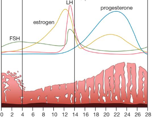
(A) During ovulation, LH surge block the ability of granulosa cells of ovarian follicle to release estrogen. This eliminates the negative feedback effect of estrogen on the hypothalamus and pituitary and further increases the release of LH. LH stimulate the rupture of the follicle and release of the oocyte from the ovary. LH stimulate granulosa cells to produce progesterone and the formation of the corpus luteum. the ruptured follicle forms the corpus luteum which has 14 days (unless the ovum is fertilized in which case the corpus luteum persists). When there is no fertilized ovum, the corpus luteum degenerates, sheds the endometrium, the corpus albicans is formed and a new cycle begins.
Hormonal changes during the menstrual cycle 5.
(C) The LH surge precedes and initiates ovulation and the release of the oocyte. The menstrual cycle describes the monthly events in female reproductive system that associated to changes in the uterus and the ovaries. These changes include endometrial shedding and the release of a mature oocyte.
During the follicular phase (the proliferative phase), the estrogen released by the follicle prior to ovulation promotes the growth and proliferation of the endometrium.
Basal body temperature is increased after ovulation under the influence of progesterone.
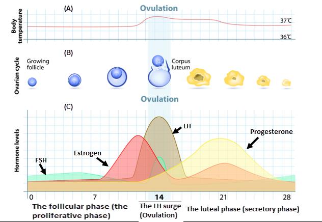
The menstrual cycle: (A) Changes in body temperature during the menstrual cycle. (B) Stages of follicular development. (C) Hormonal changes during the menstrual cycle.
6.
(D) After ovulation and during the luteal phase (secretory phase) of the menstrual cycle, the ruptured follicle forms the corpus luteum which has a duration 14 days. In the absence of fertilized ovum, the corpus luteum secrets large amounts of estrogen and progesterone, this causes the negative feedback inhibition of LH and FSH
secretion from the anterior pituitary.
The hormones produced by the ovaries and their functions Hormone
Produced by
Actions
Estrogen
Granulosa cells of the
1. Brest development
developing primordial
2. Maintain vaginal lubrication
follicles
3. Stimulate endometrial cell proliferation
4. Enhance sex drive (libido)
5. Stimulate closure of epiphyses
Progesterone Granulosa cells of the 1. Maintenance of pregnancy corpus luteum
2. Determine the woman’s fertile period by
rise in body temperature
Androgen
Theca
cells
of
the 1. Development pubic and axillary hair
ovarian follicle
2. Increase libido
Inhibin
Granulosa and theca 1. Inhibit FSH secretion cells of the ovarian 2. Role in follicle selection follicle
Activin
Granulosa cells
1. Stimulate FSH secretion
2. Development of the endometrium and
cell proliferation
Relaxin
Corpus luteum
1. Stimulate follicular development and
oocyte maturation
2. Prepare for implantation
7.
(B) The major hormones produced by the placenta is human chorionic gonadotrophic hCG. hCG is a glycoprotein resembling luteinizing hormone (LH). It is produced by the developing placenta and primary function is maintaining luteal function and preventing the regression of corpus luteum. It peaks in the first 8 weeks of pregnancy and then declines. After 8 weeks, the placenta replaces the corpus luteum as the primary organ responsible for secreting progesterone.
Placental hormones and their functions Hormones
Functions
Human
chorionic
gonadotropin Prevent the regression of corpus luteum (hCG)
and maintain pregnancy
Progesterone
After eight weeks of gestation, the placenta
begins
secreting
progesterone,
which replaces the progesterone
released from the corpus luteum.
Progesterone
increases
medullary
respiratory centres and increase
alveolar ventilation.
Estrogen
Increases
during
pregnancy
stimulate
proliferation of mammary gland.
Prevent lactation during pregnancy.
Testosterone
Increases during pregnancy. The placenta
converts androgens to estrogen due
to high aromatase activity.
Human Chorionic
Stimulate
breast
development
during
Somatomammotropin (hCS)
pregnancy
CRH
Signaling the end of pregnancy
8.
(A) This is a normal proportion of a female body fat composition. Amenorrhea is the absence of menstruation and it can be either primary amenorrhea (absence of menarche in a female by the age of 16 years) or secondary amenorrhea (disappearance of previously established menstruation).
Causes of secondary amenorrhea
Physiological
Pregnancy
Hypothalamic failure (Due to disruption Psychological stress of GnRH secretion)
Severe weight loss.
Excessive exercise.
Pituitary failure (Due to excessive Hyperprolactinemia secretion of prolactin)
Ovarian failure
Autoimmune diseases (Graves’ disease)
Genetic cause (Turner’s syndrome)
9.
(C) Menopause occurs as a result of the ovaries running out of follicles and their ability to produce estrogen declines. The lack of estrogen may produce general body changes. The levels of pituitary gonadotrophins (LH and FSH) rise markedly due to loss of estrogen negative feedback. Estrogen is required for maintenance of normal bone mass and when estrogen levels are low during menopause, this causes loss of bone mass called osteoporosis and increase incidence of bone fractures.
Body System
Symptoms
Skin
Hot flashes and night sweats.
Reproductive
Vaginal dryness, vaginal atrophy.
The uterus, ovaries and cervix (becoming
smaller and narrower).
Urinary
Increased incidence of urinary tract
infections and urinary incontinence.
Central nervous system
Anxiety, insomnia and decreased libido
Musculoskeletal
Osteoporosis, bone fracture and joint
pain.
10.
(B) There is a relationship between the placenta and the fetus known as feto-placental unit, in which the placenta produces steroid hormones such as progesterone and transfer them to the fetus. In the fetal adrenal, the progesterone is converted into precursors steroid hormone including dehydroepiandrosterone sulphate (DHEA-S) and 16-hydroxydehydroepiandrosterone sulphate (16-OH-DHEA-S). These two steroids are transported back to the placenta where they form the precursors for estrogen synthesis as human placenta has aromatase enzyme but lacks the ability to produce DHEA-S and 16-OH-DHEA-S. Thus, the formation of estrogens in the placenta is a three stages process: 1. Synthesis of progesterone in the placenta.
2. Fetal conversion of progesterone to DHEA-S and 16-OH-DHEA-S.
3. Placenta conversion of these steroids into estrogen.
1.
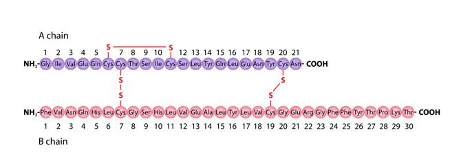
Regarding the structure of insulin, how many intermolecular and intramolecular disulfide bond is present in insulin?
A.
1 Intermolecular, 2 intramolecular
B.
2 Intermolecular, 2 intramolecular
C.
2 Intermolecular,1 intramolecular
D.
1 Intermolecular, 3 intramolecular
2.
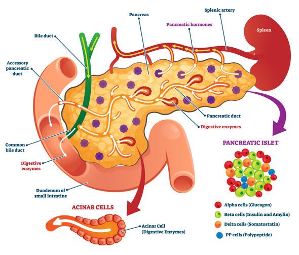
Pancreatic islets consist of specialized cells that produce different peptide hormones.
Which of the following cells produce insulin?
A.
Alpha cells.
B.
Beta cells.
C.
Gamma cells.
D.
Delta cells.
3.
Which of the following factors causes insulin secretion to increase?
A.
Increased blood glucose.
B.
Somatostatin.
C.
Fasting.
D.
Galanin.
4.
Which of the following statements about insulin secretion is FALSE?
A.
It involves closure of ATP-sensitive potassium channels.
B.
It is a biphasic process involving two pools of insulin.
C.
It involves opening of voltage-sensitive calcium channels.
D.
Glucose enters pancreatic β cells by combining with GLUT4.
5.
Which of the following statements about the insulin receptor is TRUE?
A.
Insulin receptors are linked to G proteins.
B.
Insulin binds to the β subunit on the outer surface of the cell membrane.
C.
It is a tetramer of two α and two β subunits.
D.
The binding of insulin decreases the activity of tyrosine kinase on the intracellular end of the receptor.
6.
Regarding the action of insulin in skeletal muscle, which of the following is INCORRECT?
A.
It decreases protein catabolism.
B.
It induces glycogen synthase.
C.
It decreases glucose uptake.
D.
It increases amino acid uptake.
7.
Regarding the action of insulin on adipose tissue, which of the following is INCORRECT?
A.
It induces lipoprotein lipase which actively hydrolyses triglyceride from circulating lipoproteins.
B.
It reduces circulating free fatty acids.
C.
It promotes triglyceride storage in adipocytes.
D.
Its effects appear to involve phosphorylation of lipases.
8.
A 10-year-old girl diagnosed with diabetes mellitus type 1. She asks you to explain about her condition. What is your BEST response?
A.
Your alpha cells are unable of producing and releasing insulin.
B.
The exocrine function of your pancreas should produce insulin.
C.
Without insulin, you will develop hypoglycemia.
D.
Your pancreas cannot secrete insulin.
9.
Which of the following statements regarding Glucagon is NOT true?
A.
Is produced by the beta cells of the islets of Langerhans.
B.
Is a polypeptide.
C.
Output is inversely proportional to the blood glucose level.
D.
Increases the breakdown of liver glycogen.
10.
Severe uncontrolled diabetes mellitus leads to a raised all of the following EXCEPT: A.
Urinary specific gravity and osmolality.
B.
Plasma K concentration.
C.
H+ ion concentration in body fluids.
D.
Arterial PCO2.
Answers:
1.
(C) Insulin is a peptide hormone with two chains: A and B. The A chain has 21 amino acids and the B chain has 30 amino acids. Two intermolecular disulphide bonds and one intramolecular disulphide bond hold the two-peptide chain together.
Structure of Insulin
2.
(B) The pancreas has exocrine and endocrine functions. The pancreatic acinar cells synthesize and secretes digestive enzymes directly into the small intestine (exocrine functions). Islet of Langerhans produces hormones directly into the blood (endocrine functions). The islet of Langerhans contains four types of cells: α cells which produce glucagon, β cells secrete insulin, δ cells synthesize somatostatin and PP cells secrete pancreatic polypeptide.
Hormones produced by the islet of Langerhans and their functions Hormone
Produced by
Functions
secreted
Glucagon
α cell (A cell)
Increases
blood
glucose
by
glycogenolysis and gluconeogenesis.
Insulin
β cells (B cell)
Enhance storage of glucose as glycogen.
Enhance storage of fats in adipose tissue.
Somatostatin
δ cells (D cell)
Inhibits insulin and glucagon secretion.
Pancreatic
PP cells (F cell)
polypeptide
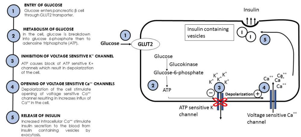
3.
(A) The most important stimulus that increases insulin secretion is an increase in the blood glucose concentration. Somatostatin, fasting and galanin inhibits insulin secretion.
Factors that stimulate or inhibit insulin secretion Increase insulin secretion
Inhibits insulin secretion
Increase blood glucose
Decreased blood glucose
Increase blood free fatty acids
Fasting
Increased blood amino acids
Somatostatin
β-Adrenergic stimulation
Galanin
Acetylcholine
α-Adrenergic stimulation
Glucagon like peptide 1 (GLP1)
Cholecystokinin
Glucose-dependent
insulinotropic
peptide (GIP)
4.
(D) The GLUT2 transporter allows glucose to enter pancreatic β cells. GLUT4 is an insulin-regulated glucose transporter protein found predominantly in skeletal muscle and adipose tissue that facilitates glucose uptake into cells.
Steps of insulin secretion from β cells of pancreas by stimulation of glucose 5.
(C) Insulin receptor is a transmembrane protein with a single transmembrane domain. It's a tetramer protein made up of two extracel ular α-subunits (where insulin binds) and two β-subunits (with tyrosine kinase activity) linked together by a disulfide bond. Insulin receptors are not linked to G protein receptors and does not act by generating second messenger but its act by intrinsic activation of tyrosine kinase.
6.
(C) Insulin is an anabolic hormone. In terms of its effect on muscle, insulin promote uptake of glucose by the muscle cells and enhances the formation of glycogen. In
terms of protein, insulin enhances protein synthesis by increasing amino acid intake and decreasing protein catabolism.
Effect of insulin’s metabolic activities on skeletal muscle Glucose metabolism
Increase uptake of glucose via GLUT4
and glycogen synthesis.
Protein metabolism
Increase uptake of amino acids, protein
synthesis and decreases protein
catabolism.
7.
(D) As an anabolic hormone, insulin increases the uptake of glucose in the adipose tissue and increases the storage of glucose as triglycerides by two mechanisms: 1.
Insulin induce the expression of the enzyme endothelial lipoprotein lipase, which releases free fatty acids and glycerol from the circulating lipoproteins and enhance the uptake of free fatty acids and glycerol by the adipocytes and thus reduce circulating free fatty acids and promote storage as triglycerides. 2. Insulin inhibits intracellular hormone sensitive lipase by decreasing the phosphorylation and thus increases esterification and storage of fatty acids.
Effect of insulin’s metabolic activities on adipose tissue Glucose metabolism
Increase uptake of glucose via GLUT4.
Protein metabolism
Increase uptake of amino acids and
protein synthesis.
Fat metabolism
Increase storage of triglycerides in
adipose tissue.
8.
(D) Type 1 diabetes mellitus (insulin dependent diabetes mellitus or IDDM) is a childhood onset. It is caused by an autoimmune process that destroys pancreatic β
cells. This leads to an excess of plasma glucose and ketones because of reduced ability of tissues to utilize glucose. Patients with untreated type 1 diabetes frequently exhibit polyuria as a result of osmotic diuresis, which happens when glucose filtration exceeds the renal transport maximum (Tm) of glucose reabsorption and thus not all the filtered glucose is reabsorbed and the glucose appears in the urine (glycosuria).
9.
(A) Glucagon a polypeptide hormone with 29-amino-acid. Produced by α cells in the pancreas. Glucagon release from pancreatic α cells is stimulated in response to low glucose levels which has the effect of increasing blood glucose levels.
Its major target is the liver, where it promotes the release of glucose.
The actions of glucagon
Tissue
Actions
Liver
Glycogenolysis – the breakdown of glycogen to glucose.
Gluconeogenesis
–
synthesis
of
glucose
from
noncarbohydrates.
Release of glucose to the blood from liver cells.
Adipose tissue Breakdown of fats (lipolysis) and raising plasma free fatty acid concentration.
(A) Insulin promote the movement of potassium into the cells. Deficiency of insulin results in hyperkalemia. In addition, the hyperglycemia and hyperosmolality leading to the movement of potassium into the extracellular fluid further contributing to hyperkalemia. Deep breathing (Kussmaul’s breathing) respiration develops in an attempt to compensate for metabolic acidosis and decreased pH. This decreases the partial pressure of carbon dioxide (PCO2).
insulin production
No glucose uptake by cells
(Hyperglycemia)
Glucagon
Lipolysis
Muscle breakdown
Liver
Oxidation of fatty acids
Glycogenolysis and
and production of ketone bodies
Gluconeogenesis
Acetoacetate
Beta Hydroxybutyrate
Hyperglycemia
Acetone
Reduce pH
Plasma Osmolality Hyperosmolality
Hyperkalemia
Kussmaul’s respiration
Hyperglycemia exceeds
Renal threshold
Osmotic diuresis
Reduce blood volume
Glycosuria
Polyuria
Consequences of insulin deficiency
1.
Calcium level in the blood is regulated by the A.
Parathyroid, kidney and thyroid
B.
Adrenal medulla, skin and pancreas
C.
Testes, hypothalamus and liver
D.
Parathyroid, thymus and heart
2.
What stimulates the release of PTH from the parathyroid gland?
A.
TSH from the posterior pituitary gland
B.
high levels of calcium in the blood
C.
calcitonin from the anterior pituitary gland
D.
low levels of calcium in the blood
3.
All the following actions of parathyroid hormone is true EXCEPT: A.
Secretion is regulated by a hypothalamus feedback control system.
B.
Acts directly on bone to increase bone resorption.
C.
Increases phosphate excretion.
D.
Promotes absorption of calcium from the intestines.
4.
Which of the following is TRUE regarding the concentration of ionized calcium in plasma?
A.
Less than the free ionized calcium concentration in intracellular fluid.
B.
The main regulator of parathyroid hormone secretion.
C.
About 65 per cent of the total plasma calcium concentration.
D.
Reduced when the plasma protein level rises.
5.
The hormonal actions on bone includes the following EXCEPT: A.
Growth hormone stimulate bone growth during childhood.
B.
Parathyroid hormone decreases bone resorption.
C.
Glucocorticoids promote bone resorption.
D.
Estrogen stimulate the epiphyses of long bones to unite.
6.
Regarding the effect of vitamin D on plasma calcium concentration and the bone includes all the following EXCEPT:
A.
It increases renal tubular calcium and phosphate reabsorption.
B.
It causes an increase in synthesis of calcium-binding protein.
C.
It decreases bone formation.
D.
It increases intestinal calcium and phosphate absorption.
7.
Which of the following best describes vitamin D?
A.
It is a water-soluble vitamin.
B.
Deficiency is seen in areas with high sun exposure.
C.
Calcitriol is the physiologically active form of vitamin D.
D.
It is converted to 1,25-dihydroxycholecalciferol in the liver.
All the following conditions are associated with vitamin D deficiency EXCEPT: A.
Osteomalacia.
B.
Hyperparathyroidism.
C.
Rickets.
D.
Kidney stones.
9.
Which of the following profiles for plasma calcium (Ca2+), phosphate (P) and parathyroid hormone (PTH) concentration would be expected in a patient with chronic renal failure?
Plasma (Ca2+)
Plasma (P)
Plasma (PTH)
A.
Low
Low
Low
B.
High
Low
Low
C.
Low
High
High
D.
High
High
High
10.
Which of the following statements about calcitonin is FALSE?
A.
It stimulates the number and the activity of osteoclast.
B.
It reduces blood calcium levels.
C.
It is produced by the parafollicular cells of the thyroid gland.
D.
It is secretion occurs when the blood phosphate level rises.
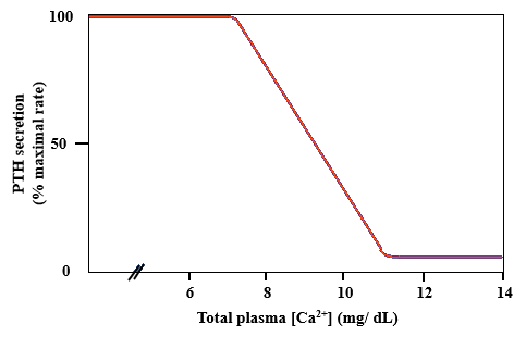
Answers:
1.
(A) Calcium homeostasis involves THREE systems (bone, kidney and GI tract) and THREE hormones (parathyroid hormone, calcitonin and vitamin D) Sources of calcium are dietary (absorbed in the gut), reabsorption of calcium by the kidney and bone.
Hormonal control of serum calcium
Gland
Hormone
Produced by
Parathyroid gland Parathyroid hormone
Chief cells of parathyroid gland
Kidney
Vitamin D (Calcitriol)
Proximal convoluted tubule of kidney
Thyroid gland
Calcitonin
Parafollicular cells of thyroid gland
2.
(D) PTH secretion is regulated by the plasma Ca++ concentration. When the total Ca concentration is in the normal range (i.e., 10 mg/dL) or higher, PTH is secreted at a low (basal) level. However, when the plasma Ca++ concentration decreases to less than 10 mg/dL, PTH secretion is stimulated, reaching maximal rates when the Ca++
concentration is 7.5 mg/dL. The response of the parathyroid glands to a decrease in ionized Ca++ concentration is remarkably prompt, occurring within seconds.
Furthermore, the faster the ionized Ca++ falls, the greater the PTH secretory response.
Plasma Calcium Concentration and PTH Secretion: The ionized Ca2+
concentration regulates secretion by the parathyroid glands. The PTH
secretion reach the maximum when the calcium concentration is below 8
mg/dL.
3.
(A) The hormones secreted by the parathyroid glands and the pancreas are not under the influence of the releasing hormones secreted by hypothalamus and the pituitary gland. The parathyroid hormone is regulated directly by the calcium level in the blood.
Actions of parathyroid hormone on the bone:
The overall effect of PTH on bone is to promote bone resorption, delivering both Ca and phosphate to the extracellular fluid (ECF). The phosphate released from bone will complex with Ca++ in ECF and limit the rise in ionized Ca++ concentration. Thus, an additional mechanism must coordinate with the PTH effect on bone to cause the plasma ionized Ca++ concentration to increase. (That additional mechanism is the phosphaturic action of PTH.).
Actions of parathyroid hormone on kidney:
(1) PTH inhibits phosphate reabsorption by inhibiting Na+-phosphate co-transport in the proximal convoluted tubule. As a result of this action, PTH causes phosphaturia, an increased excretion of phosphate in urine.
(2) PTH stimulates Ca++ reabsorption. This second renal action of PTH is on the distal convoluted tubule and complements the increase in plasma Ca++ concentration that resulted from the combination of bone resorption and phosphaturia.
Actions of parathyroid hormone on Gastrointestinal system: PTH is indirectly acts on the gastrointestinal tract by stimulating 1,25-dihydroxycholecalciferol production.
Parathyroid hormone
Bone
GIT
Kidney
PTH increases Ca
PTH increases
1. Parathyroid hormone increases the
absorption. Indirect via
bone resorption
reabsorption of calcium from the
formation of 1,25-
distal convoluted tubule.
dihydroxycholecalciferol
(Calcitriol)
2. Parathyroid hormone activates the
synthesis of vitamin D from the
proximal convoluted tubule.
3. Parathyroid hormone increases the
excretion of phosphate.
Parathyroid hormone increases serum calcium
Actions of parathyroid hormone (PTH) on the bone, GIT and kidneys 4.
(B) PTH secretion is the main regulator of plasma Ca++ concentration.
Total Ca concentration in blood is normally kept at 10 mg/dL, which is equivalent to 2.5 mmol/L. The calcium in the Plasma is available in three forms: 1. About 41% is bound to plasma proteins (1 mmol/L).
2. About 9% complexed to anions (phosphate, citrate, sulfate) (0.2 mmol/L).
3. The remaining 50% is free, ionized – only this fraction is biologically active (1.2
mmol/L) or (2.4 mEq/L).
Reduced plasma proteins will lower the protein-bound calcium not ionized calcium.
The concentration of intracellular calcium is about 0.0001 mM approximately 10,000
times lower than that of the extracellular space.
5.
(B) The regulation of bone is affected through the actions of several hormones.
Parathyroid hormone promotes bone resorption by initially stimulating the bone forming cells the osteoblast to express RANKL (receptor activator of NF-KB ligand) which binds to RANK receptor on the cells responsible for bone resorption the osteoclasts which secrete an enzyme called acid protease to breakdown the extracellular collagen matrix.
The hormonal actions on bone
Hormone
Action
Parathyroid hormone (PTH)
Stimulate bone resorption
Calcitriol
Maintain osteoblast functions.
Glucocorticoids
Promote bone resorption by stimulating osteoclast activity
Estrogen
Promote bone formation by fusion of epiphysis with the shaft of the bone
Testosterone
Promote bone formation by fusion of epiphysis with the shaft of the bone
Growth hormone
Stimulate bone growth by promoting proliferation of cartilage cells in the epiphyseal plate
Insulin-like growth factor 1 Stimulate bone growth by promoting proliferation of (IGF-1)
cartilage cells in the epiphyseal plate
6.
(C) Vitamin D promote bone structure by stimulating osteoblast functions Effects of vitamin D (calcitriol) on plasma calcium and bone System
Effects
Gastrointestinal tract
Increase uptake of calcium from the gut by stimulating the synthesis of calbindin protein that binds calcium to increase its absorption.
Kidney
Decreasing urinary excretion of calcium by increasing renal tubular calcium reabsorption.
Bone
Maintain bone structure by stimulating osteoblast action.
7.
(C) Two sources are available for vitamin D. The first is vitamin D3 (cholecalciferol) which can be either produced in the skin when exposed to UV radiation, which facilitated by cholesterol metabolism or obtained through dietary sources and the second is vitamin D2 (ergocalciferol) which is derived from yeast and fungi as food supplement. Both sources are activated in the kidneys to form calcitriol.
Dietary Intake
Dietary Intake
Ultraviolet light
Fish/Meat/Liver
(Supplement)
Cholecalciferol
(Vitamin D3)
Ergocalciferol
(Vitamin D2)
Liver
25-Hydroxycholecalciferol
Kidney
1,25-Dihydroxycolecalciferol
(Calcitriol)
Bone
Small intestine
Kidney
Maintain osteoblast
Increases calcium phosphate
Reduces calcium
functions
absorption
phosphate excretion
Sources, activation and functions of vitamin D. Two sources of vitamin D, from the skin through the exposure to sunlight and dietary intake. Activation of vitamin D
require two hydroxylation steps, the first in the liver and the second in the kidney.
Two stimuli are essential for the synthesis of vitamin D, parathyroid hormone (PTH) and low phosphate levels.
8.
(D) Vitamin D is essential hormone that regulate calcium concentration by increasing the intestinal uptake of calcium from the diet and maintain normal calcification of bones by stimulating the actions of osteoblast. The potential causes for vitamin D
deficiency include:
1. Inadequate dietary intake of calcium from animal protein sources and dairy products.
2. Inadequate exposure to sunlight.
3. Chronic renal failure as the damaged kidney is not able to produce vitamin D.
The consequences of vitamin D deficiency results in osteomalacia (reduced mineralization of bones in adult), rickets (reduced mineralization of bones in children) and compensatory increase in parathyroid hormone to maintain the normal level of plasma calcium.
Excess circulating of vitamin D causes kidney stones, polyuria and an increase in plasma calcium concentration (hypercalcemia).
9.
(C) A decrease in the glomerular filtration rate (GFR) seen in renal failure leads to a decrease in filtration of phosphate, which in turn causes phosphate retention and an increase in plasma phosphate concentration (Hyperphosphatemia). The excess phosphate then reacts with the ionized calcium as shown in the following reaction and decreases the plasma calcium concentration (Hypocalcemia).
Ca2+ + HPO 2-
4
CaHPO4
Reduction in calcium concentration will stimulate the release of parathyroid hormone (PTH) as an attempt to increase blood calcium level and increase excretion of phosphate. As a result, secondary hyperparathyroidism develops, which in turn leads to a significant bone resorption and bone disease called osteitis fibrosa cystica, development of metastatic calcification and osteomalacia. These bone diseases manifestation which is due to renal failure collectively called renal osteodystrophy.
10.
(D) Calcitonin is synthesized and secreted by the parafollicular or C-cells of the thyroid gland. The major action of calcitonin is to inhibit osteoclastic bone resorption which decreases the plasma calcium concentration and decreases the reabsorption of phosphate and calcium from the kidney. The net effect will be to reduce both plasma calcium and phosphate concentration.
The major stimulus for calcitonin secretion is increased plasma concentration.
In contrast to PTH, calcitonin does not participate in the minute-to-minute regulation of the plasma calcium concentration. The physiologic role for calcitonin in is uncertain because neither thyroidectomy (with decreased calcitonin) nor thyroid tumors (with increased calcitonin levels) cause a derangement of calcium metabolism, as would be expected if calcitonin had important regulatory functions.
It has been suggested that calcitonin may have a minor role in protecting the bones of the mother during pregnancy and lactation.
1.
Which of the following is a mitochondrial enzyme that mediates all steps in the conversion of cholesterol to pregnenolone and is the rate limiting step for the entire steroid synthetic pathway?
A.
Cholesterol side chain cleavage
B.
17α-hydroxylase
C.
21 hydroxylase
D.
11β-hydroxylase
2.
Which of the following adrenal medulla's enzymes converts norepinephrine into epinephrine?
A.
Tyrosine hydroxylase
B.
Phenylethanolamine N-methyltransferase (PNMT)
C.
Amino acid decarboxylase
D.
Phenylalanine hydroxylase.
3.
Which of the following statements about melatonin is CORRECT?
A.
Is produced mainly in the anterior lobe of the pituitary gland.
B.
The secretion of melatonin is controlled by environmental light and dark periods.
C.
Melatonin secretion stimulate appetite.
D.
The secretion of melatonin is stimulated by exposure to light.
4.
Selective destruction of the zona fasciculata of the adrenal cortex would produce a deficiency of which hormone?
A.
Aldosterone.
B.
Cortisol.
C.
Testosterone
D.
Adrenaline.
5.
All the following conditions increases erythropoietin production from the kidney EXCEPT:
A.
Chronic renal failure.
B.
Obstructive lung diseases.
C.
Hemolytic anemia.
D.
Low arterial blood partial pressure of oxygen (PO2).
6.
Which of the following gastrointestinal hormone is most important in insulin secretion?
A.
Vasoactive intestinal polypeptide (VIP).
B.
Gastrin releasing peptide (GRP).
C.
Substance P.
D.
Gastric inhibitory peptide (GIP).
7.
Which of the following gastrointestinal hormone is involved in the secretion of enzymatic components of pancreatic juice?
A.
Gastrin.
B.
Secretin.
C.
Cholecystokinin
D.
Motilin
Which of the following conditions are the MOST likely to lead to the release of atrial natriuretic peptide (ANP)?
A.
High blood volume.
B.
Low blood volume.
C.
Low blood pressure.
D.
Hyponatremia.
9.
Which of two hormones act on the hypothalamus to increase satiety and hunger, respectively?
A.
Leptin and insulin.
B.
Ghrelin and orexin.
C.
Insulin and peptide YY.
D.
Leptin and ghrelin.
10.
Which of the following hormones requires the amino acid tryptophan for its synthesis?
A.
Melatonin and serotonin.
B.
Thyroxine and triiodothyronine.
C.
Estrogen and progesterone.
D.
Cortisol and cortisone.
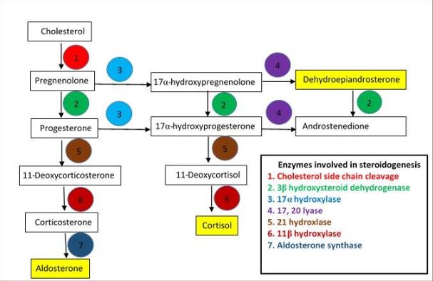
Answers:
1.
(A) Cholesterol side chain cleavage (CSCC) mediates all steps in the conversion of cholesterol to pregnenolone and is the rate limiting step for the entire steroid synthetic pathway.
Pathways of steroid hormone synthesis in the adrenal cortex. Seven different enzymes involved in the formation of steroid hormones (aldosterone, cortisol and DHEA)
2.
(B) The major hormone of adrenal medulla is adrenaline, whereas noradrenalin is more abundant as neurotransmitter of the central and sympathetic nervous system.
Dopamine is secreted in small amounts.
The synthesis of catecholamine is initiated by conversion of tyrosine to dihydroxyphenylalanine (DOPA) by the enzyme tyrosine hydroxylase (TH). Then it is decarboxylated by amino acid decarboxylase to form dopamine. Dopamine is then converted to noradrenaline by dopamine β-hydroxylase (DBH). The final step is the conversion
of
noradrenaline
to
adrenaline
by
phenylethanolamine-N-
methyltransferase (PNMT).
Tyrosine
Tyrosine hydroxylase
Dihydroxyphenylalanine
(DOPA)
Amino acid decarboxylase
Dopamine
Dopamine β-hydroxylase
Noradrenaline
Phenylethanolamine N-methyl transferase
(PNMT)
Adrenaline
Synthesis of adrenal medulla hormones
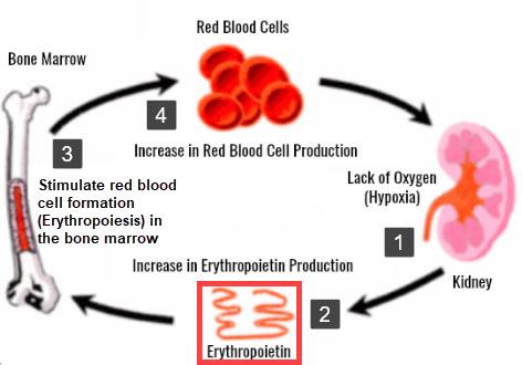
3.
(B) Melatonin is a hormone produced in the pineal gland which involved in regulation of circadian rhythms. Melatonin production is highest during the night-time and negatively controlled by exposure to light.
4.
(B) Cortisol is produced from the adrenal’s cortex zona fasciculata in response to stress. Autoimmune destruction of the adrenal glands results in insufficient production of cortisol as seen in Addison's disease. The clinical features of adrenal insufficiency:
• Weakness and lethargy.
• Hyponatremia decreased reabsorption of sodium and water.
• Hyperkalemia increased potassium concentration.
• Hypotension and hypovolemia due to excessive excretion of sodium.
5.
(A) In chronic renal failure, the kidney produces less erythropoietin due to damage to the erythropoietin producing cells.
Formation and action of erythropoietin
Erythropoietin is a peptide hormone.
Produced by the fibroblast cells in the
peritubular interstitium of the kidney.
Erythropoietin promotes red blood cell
(RBC) formation from the bone marrow
in response to low partial pressure of
oxygen (PO2).
6.
(D) Gastric inhibitory peptide (GIP) is a peptide hormone synthesized from the K
cells in the duodenum and jejunum. It is major function is to induce insulin secretion.
Gastrointestinal hormones site of production and their actions Hormone
Produced by
Actions
GIP
K cells in duodenum and Increases insulin secretion jejunum
VIP
Myenteric and submucosal Induce relaxation of GI smooth neurons
of
the
enteric muscle and sphincter relaxation
nervous system in the GI
GRP
Vagal nerve ending that Stimulate the release of gastrin innervate the G cells of the from the stomach stomach
Substance P Myenteric and submucosal Increases motility of the small neurons
of
the
enteric intestine
nervous system in the GI
7.
(C) Cholecystokinin stimulates the release of pancreatic enzymatic, stimulates contraction of the gall bladder, relaxation of the sphincter of Oddi and inhibits gastric secretions.
Gastrointestinal hormones site of production and their actions Hormone
Produced by
Actions
Gastrin
G cells in antrum of stomach
Promote gastric acid
secretion
Secretin
S cells, in the duodenum and Increases bicarbonate jejunum
secretion
from
the
pancreas
Cholecystokinin (CCK)
I cell in duodenum
Increases pancreatic
enzyme
Motilin
M cells, in the duodenum Increases contraction and jejunum
of smooth muscle in
the
stomach
and
duodenum
and
promote GI motility
8.
(A) Atrial natriuretic peptide (ANP) is a hormone produced in the heart mainly from the right atrium in response to high blood volume. ANP inhibits the reabsorption of sodium in the nephron’s collecting duct through closing the luminal membrane sodium channels and thus increases urinary excretion of sodium and water. It is a vasodilator which lowers the systemic blood pressure. The action of ANP is opposes the actions of aldosterone which produced in response to low blood volume that stimulate the reabsorption and sodium retention.