Help Me Understand Genetics by NIH - HTML preview
Download the book in PDF, ePub for a complete version.
Handbook
Help Me Understand Genetics
Reprinted from Genetics Home Reference (http://ghr.nlm.nih.gov/) Lister Hill National Center for Biomedical Communications
U.S. National Library of Medicine
National Institutes of Health
Department of Health & Human Services
Published November 12, 2013
Genetics Home Reference - http://ghr.nlm.nih.gov/
Handbook
Handbook
Table of Contents
Cells and DNA
Cells, genes, and chromosomes
How Genes Work
Proteins, cell growth, and cell division
Mutations and Health
Gene mutations, chromosomal changes, and conditions that run
in families
Inheriting Genetic Conditions
Inheritance patterns and understanding risk
Genetic Consultation
Finding and visiting a genetic counselor or other genetics
professional
Genetic Testing
Benefits, costs, risks, and limitations of genetic testing
Gene Therapy
Experimental techniques, safety, ethics, and availability
The Human Genome Project
Sequencing and understanding the human genome
Genomic Research
Next steps in studying the human genome
page 2
Genetics Home Reference - http://ghr.nlm.nih.gov/
Handbook
Cells and DNA
Chapter 1
Cells and DNA
Table of Contents
What is a cell?
What is DNA?
What is mitochondrial DNA?
What is a gene?
What is a chromosome?
How many chromosomes do people have?
page 3
Genetics Home Reference - http://ghr.nlm.nih.gov/
Handbook
Cells and DNA
What is a cell?
Cells are the basic building blocks of all living things. The human body is composed
of trillions of cells. They provide structure for the body, take in nutrients from food,
convert those nutrients into energy, and carry out specialized functions. Cells also
contain the body’s hereditary material and can make copies of themselves.
Cells have many parts, each with a different function. Some of these parts, called
organelles, are specialized structures that perform certain tasks within the cell.
Human cells contain the following major parts, listed in alphabetical order:
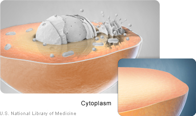
Cytoplasm (illustration on page 5)
Within cells, the cytoplasm is made up of a jelly-like fluid (called the cytosol)
and other structures that surround the nucleus.
Cytoskeleton
The cytoskeleton is a network of long fibers that make up the cell’s structural
framework. The cytoskeleton has several critical functions, including
determining cell shape, participating in cell division, and allowing cells to move.
It also provides a track-like system that directs the movement of organelles
and other substances within cells.
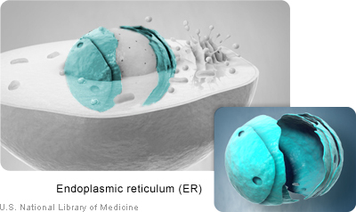
Endoplasmic reticulum (ER) (illustration on page 6)
This organelle helps process molecules created by the cell. The endoplasmic
reticulum also transports these molecules to their specific destinations either
inside or outside the cell.
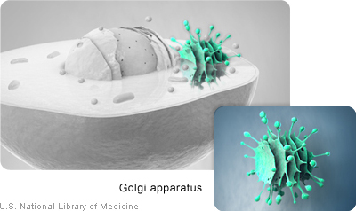
Golgi apparatus (illustration on page 6)
The Golgi apparatus packages molecules processed by the endoplasmic
reticulum to be transported out of the cell.
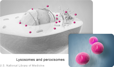
Lysosomes and peroxisomes (illustration on page 6)
These organelles are the recycling center of the cell. They digest foreign
bacteria that invade the cell, rid the cell of toxic substances, and recycle
worn-out cell components.
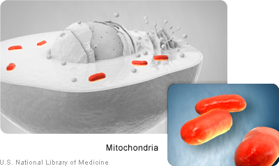
Mitochondria (illustration on page 7)
Mitochondria are complex organelles that convert energy from food into a
form that the cell can use. They have their own genetic material, separate
from the DNA in the nucleus, and can make copies of themselves.
page 4
Genetics Home Reference - http://ghr.nlm.nih.gov/
Handbook
Cells and DNA
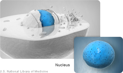
Nucleus (illustration on page 7)
The nucleus serves as the cell’s command center, sending directions to the
cell to grow, mature, divide, or die. It also houses DNA (deoxyribonucleic acid),
the cell’s hereditary material. The nucleus is surrounded by a membrane called
the nuclear envelope, which protects the DNA and separates the nucleus from
the rest of the cell.
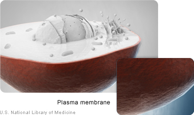
Plasma membrane (illustration on page 7)
The plasma membrane is the outer lining of the cell. It separates the cell from
its environment and allows materials to enter and leave the cell.
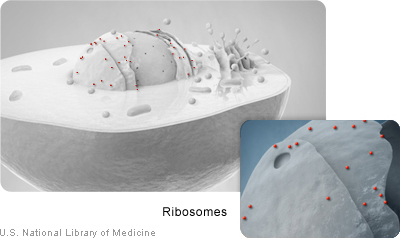
Ribosomes (illustration on page 8)
Ribosomes are organelles that process the cell’s genetic instructions to create
proteins. These organelles can float freely in the cytoplasm or be connected
to the endoplasmic reticulum (see above).
For more information about cells:
The Genetic Science Learning Center at the University of Utah offers an interactive
introduction to cells (http://learn.genetics.utah.edu/content/begin/cells/) and their many functions.
Additional information about the cytoskeleton, including an illustration, is available
from the Cytoplasm Tutorial (http://www.biology.arizona.edu/Cell_bio/tutorials/
cytoskeleton/page1.html). This resource is part of The Biology Project at the
University of Arizona.
Illustrations
The cytoplasm surrounds the cell’s nucleus and organelles.
page 5
Genetics Home Reference - http://ghr.nlm.nih.gov/
Handbook
Cells and DNA
The endoplasmic reticulum is involved in molecule processing and transport.
The Golgi apparatus is involved in packaging molecules for export from the
cell.
Lysosomes and peroxisomes destroy toxic substances and recycle worn-out
cell parts.
page 6
Genetics Home Reference - http://ghr.nlm.nih.gov/
Handbook
Cells and DNA
Mitochondria provide the cell’s energy.
The nucleus contains most of the cell’s genetic material.
The plasma membrane is the outer covering around the cell.
page 7
Genetics Home Reference - http://ghr.nlm.nih.gov/
Handbook
Cells and DNA
Ribosomes use the cell’s genetic instructions to make proteins.
page 8
Genetics Home Reference - http://ghr.nlm.nih.gov/
Handbook
Cells and DNA
What is DNA?
DNA, or deoxyribonucleic acid, is the hereditary material in humans and almost all
other organisms. Nearly every cell in a person’s body has the same DNA. Most
DNA is located in the cell nucleus (where it is called nuclear DNA), but a small
amount of DNA can also be found in the mitochondria (where it is called
mitochondrial DNA or mtDNA).
The information in DNA is stored as a code made up of four chemical bases: adenine
(A), guanine (G), cytosine (C), and thymine (T). Human DNA consists of about 3
billion bases, and more than 99 percent of those bases are the same in all people.
The order, or sequence, of these bases determines the information available for
building and maintaining an organism, similar to the way in which letters of the
alphabet appear in a certain order to form words and sentences.
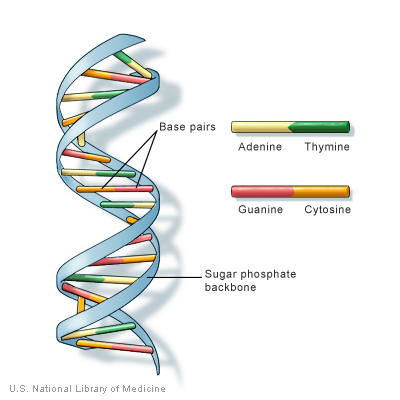
DNA bases pair up with each other, A with T and C with G, to form units called base
pairs. Each base is also attached to a sugar molecule and a phosphate molecule.
Together, a base, sugar, and phosphate are called a nucleotide. Nucleotides are
arranged in two long strands that form a spiral called a double helix. The structure
of the double helix is somewhat like a ladder, with the base pairs forming the ladder’s
rungs and the sugar and phosphate molecules forming the vertical sidepieces of
the ladder.
An important property of DNA is that it can replicate, or make copies of itself. Each
strand of DNA in the double helix can serve as a pattern for duplicating the sequence
of bases. This is critical when cells divide because each new cell needs to have an
exact copy of the DNA present in the old cell.
page 9
Genetics Home Reference - http://ghr.nlm.nih.gov/
Handbook
Cells and DNA
DNA is a double helix formed by base pairs attached to a sugar-phosphate
backbone.
For more information about DNA:
The National Human Genome Research Institute fact sheet Deoxyribonucleic Acid
(DNA) (http://www.genome.gov/25520880) provides an introduction to this molecule.
Information about the genetic code (http://geneed.nlm.nih.gov/topic_subtopic.php?
tid=15&sid=19) and the structure of the DNA double helix (http://geneed.nlm.nih.gov/
topic_subtopic.php?tid=15&sid=16) is available from GeneEd.
The New Genetics, a publication of the National Institute of General Medical
Sciences, discusses the structure of DNA and how it was discovered
(http://publications.nigms.nih.gov/thenewgenetics/chapter1.html#c1).
page 10
Genetics Home Reference - http://ghr.nlm.nih.gov/
Handbook
Cells and DNA
What is mitochondrial DNA?
Although most DNA is packaged in chromosomes within the nucleus, mitochondria
also have a small amount of their own DNA. This genetic material is known as
mitochondrial DNA or mtDNA.
Mitochondria (illustration on page 7) are structures within cells that convert the energy from food into a form that cells can use. Each cell contains hundreds to
thousands of mitochondria, which are located in the fluid that surrounds the nucleus
(the cytoplasm).
Mitochondria produce energy through a process called oxidative phosphorylation.
This process uses oxygen and simple sugars to create adenosine triphosphate
(ATP), the cell’s main energy source. A set of enzyme complexes, designated as
complexes I-V, carry out oxidative phosphorylation within mitochondria.
In addition to energy production, mitochondria play a role in several other cellular
activities. For example, mitochondria help regulate the self-destruction of cells
(apoptosis). They are also necessary for the production of substances such as
cholesterol and heme (a component of hemoglobin, the molecule that carries oxygen
in the blood).
Mitochondrial DNA contains 37 genes, all of which are essential for normal
mitochondrial function. Thirteen of these genes provide instructions for making
enzymes involved in oxidative phosphorylation. The remaining genes provide
instructions for making molecules called transfer RNAs (tRNAs) and ribosomal
RNAs (rRNAs), which are chemical cousins of DNA. These types of RNA help
assemble protein building blocks (amino acids) into functioning proteins.
For more information about mitochondria and mitochondrial DNA:
Molecular Expressions, a web site from the Florida State University Research
Foundation, offers an illustrated introduction to mitochondria and mitochondrial
DNA (http://micro.magnet.fsu.edu/cells/mitochondria/mitochondria.html).
An overview of mitochondrial DNA (http://neuromuscular.wustl.edu/mitosyn.html#
general) is available from the Neuromuscular Disease Center at Washington
University.
page 11
Genetics Home Reference - http://ghr.nlm.nih.gov/
Handbook
Cells and DNA
What is a gene?
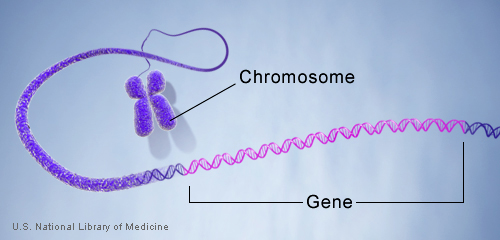
A gene is the basic physical and functional unit of heredity. Genes, which are made
up of DNA, act as instructions to make molecules called proteins. In humans, genes
vary in size from a few hundred DNA bases to more than 2 million bases. The
Human Genome Project has estimated that humans have between 20,000 and
25,000 genes.
Every person has two copies of each gene, one inherited from each parent. Most
genes are the same in all people, but a small number of genes (less than 1 percent
of the total) are slightly different between people. Alleles are forms of the same
gene with small differences in their sequence of DNA bases. These small differences
contribute to each person’s unique physical features.
Genes are made up of DNA. Each chromosome contains many genes.
For more information about genes:
Genetics Home Reference provides consumer-friendly gene summaries
(http://ghr.nlm.nih.gov/BrowseGenes) that include an explanation of each gene’s
normal function and how mutations in the gene cause particular genetic conditions.
The Centre for Genetics Education offers a fact sheet that introduces genes and
chromosomes (http://www.genetics.edu.au/Information/Genetics-Fact-Sheets/
Genes-and-Chromosomes-FS1).
page 12
Genetics Home Reference - http://ghr.nlm.nih.gov/
Handbook
Cells and DNA
What is a chromosome?
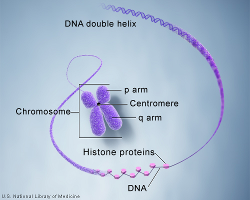
In the nucleus of each cell, the DNA molecule is packaged into thread-like structures
called chromosomes. Each chromosome is made up of DNA tightly coiled many
times around proteins called histones that support its structure.
Chromosomes are not visible in the cell’s nucleus—not even under a
microscope—when the cell is not dividing. However, the DNA that makes up
chromosomes becomes more tightly packed during cell division and is then visible
under a microscope. Most of what researchers know about chromosomes was
learned by observing chromosomes during cell division.
Each chromosome has a constriction point called the centromere, which divides
the chromosome into two sections, or “arms.” The short arm of the chromosome is
labeled the “p arm.” The long arm of the chromosome is labeled the “q arm.” The
location of the centromere on each chromosome gives the chromosome its
characteristic shape, and can be used to help describe the location of specific
genes.
DNA and histone proteins are packaged into structures called chromosomes.
page 13
Genetics Home Reference - http://ghr.nlm.nih.gov/
Handbook
Cells and DNA
For more information about chromosomes:
Genetics Home Reference provides information about each human chromosome
(http://ghr.nlm.nih.gov/chromosomes) written in lay language.
The Centre for Genetics Education offers a fact sheet that introduces genes and
chromosomes (http://www.genetics.edu.au/Information/Genetics-Fact-Sheets/
Genes-and-Chromosomes-FS1).
GeneEd also provides information about the basics of chromosomes
(http://geneed.nlm.nih.gov/topic_subtopic.php?tid=15&sid=17).
page 14
Genetics Home Reference - http://ghr.nlm.nih.gov/
Handbook
Cells and DNA
How many chromosomes do people have?
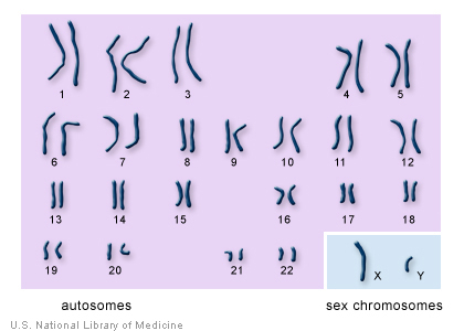
In humans, each cell normally contains 23 pairs of chromosomes, for a total of 46.
Twenty-two of these pairs, called autosomes, look the same in both males and
females. The 23rd pair, the sex chromosomes, differ between males and females.
Females have two copies of the X chromosome, while males have one X and one
Y chromosome.
The 22 autosomes are numbered by size. The other two chromosomes, X and
Y, are the sex chromosomes. This picture of the human chromosomes lined
up in pairs is called a karyotype.
For more information about the 23 pairs of human chromosomes:
Genetics Home Reference provides information about each human chromosome
(http://ghr.nlm.nih.gov/chromosomes) written in lay language.
page 15
Genetics Home Reference - http://ghr.nlm.nih.gov/
Handbook
How Genes Work
Chapter 2
How Genes Work
Table of Contents
What are proteins and what do they do?
How do genes direct the production of proteins?
Can genes be turned on and off in cells?
What is the epigenome?
How do cells divide?
How do genes control the growth and division of cells?
How do geneticists indicate the location of a gene?
What are gene families?
page 16
Genetics Home Reference - http://ghr.nlm.nih.gov/
Handbook
How Genes Work
What are proteins and what do they do?
Proteins are large, complex molecules that play many critical roles in the body.
They do most of the work in cells and are required for the structure, function, and
regulation of the body’s tissues and organs.
Proteins are made up of hundreds or thousands of smaller units called amino acids,
which are attached to one another in long chains. There are 20 different types of
amino acids that can be combined to make a protein. The sequence of amino acids
determines each protein’s unique 3-dimensional structure and its specific function.
Proteins can be described according to their large range of functions in the body,
listed in alphabetical order:
Examples of protein functions
Function
Description
Example
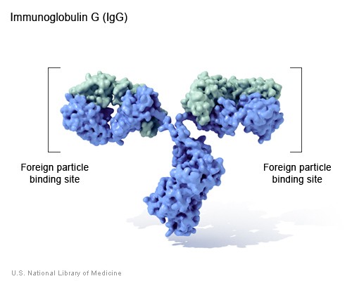
Antibody
Antibodies bind to specific foreign particles, Immunoglobulin G
such as viruses and bacteria, to help
(IgG)
protect the body.
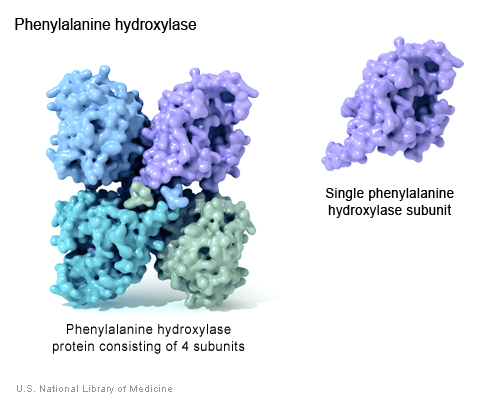
Enzyme
Enzymes carry out almost all of the
Phenylalanine
thousands of chemical reactions that take hydroxylase
place in cells. They also assist with the
formation of new molecules by reading the
genetic information stored in DNA.
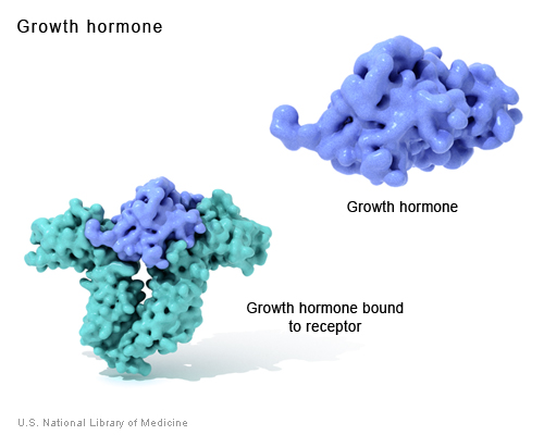
Messenger
Messenger proteins, such as some types Growth hormone
of hormones, transmit signals to coordinate (illustration on page 20) biological processes between different
cells, tissues, and organs.
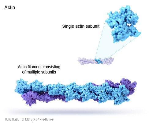
Structural
These proteins provide structure and
Actin
component
support for cells. On a larger scale, they
also allow the body to move.
Transport/storage
These proteins bind and carry atoms and Ferritin
small molecules within cells and throughout (illustration on page 22) the body.
For more information about proteins and their functions:
KidsHealth from Nemours offers a basic overview of proteins (http://kidshealth.org/
kid/stay_healthy/body/protein.html) and what they do.
page 17
Genetics Home Reference - http://ghr.nlm.nih.gov/
Handbook
How Genes Work
Illustrations
Immunoglobulin G is a type of antibody that circulates in the blood and
recognizes foreign particles that might be harmful.
page 18
Genetics Home Reference - http://ghr.nlm.nih.gov/
Handbook
How Genes Work
The functional phenylalanine hydroxylase enzyme is made up of four identical
subunits. The enzyme converts the amino acid phenylalanine to another amino
acid, tyrosine.
page 19
Genetics Home Reference - http://ghr.nlm.nih.gov/
Handbook
How Genes Work
Growth hormone is a messenger protein made by the pituitary gland. It regulates
cell growth by binding to a protein called a growth hormone receptor.
page 20
Genetics Home Reference - http://ghr.nlm.nih.gov/
Handbook
How Genes Work
Actin filaments, which are structural proteins made up of multiple subunits