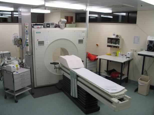
hence, the term "axial." The machine sends out a thin X-ray beam at 160 different points. Crystals
positioned at the opposite points of the beam pick up and record the absorption rates of the varying thicknesses of tissue and bone. The data are then relayed to a computer that turns the information into a
2-dimensional cross-sectional image. Risks
CT scan risks are similar to those of conventional X-rays. During the CT scan, you're briefly
exposed to radiation. But scientists believe that CT scans provide enough valuable information to
outweigh the associated risks. But if the subject or the patient: Pregnant it may be recommended to do
another type of exam to reduce the possible risk of exposing his fetus to radiation. Have asthma or
allergies. And the CT scan requires a contrast medium, there's a slight risk of an allergic reaction to the
contrast medium. Have certain medical conditions. Diabetes, asthma, heart disease, kidney problems or
thyroid conditions also increase the risk of a reaction to contrast medium.
MRI
Although CAT scanning was a breakthrough, in many
cases it was substituted by Magnetic resonance imaging
(also known as MRI) since magnetic resonance imaging is
a method of looking inside the body without using x-rays,
harmful dyes or surgery. Instead, radio waves and a strong
magnetic field are used in order to provide remarkably
clear and detailed pictures of internal organs and tissues.
History and Development of MRI
A full size MRI-Scanner. ( GFDL – Kasuga Huang)
MRI is based on a physics phenomenon, called nuclear
magnetic resonance (NMR), which was discovered in1930s by
Felix Bloch (working at Stanford University) and Edward
Purcell(from Harvard University). In this resonance, magnetic fields and radio waves cause atoms to give off tiny radio
signals. In the year 1970, Raymond Damadian, a medical
doctor and research scientist, discovered the basis for using
magnetic resonance imaging as a tool for medical diagnosis.
Four years later a patent was granted, which was the worlds
first patent issued in the field of MRI. In 1977, Dr. Damadian
completed the construction of the first “whole-body” MRI
scanner, which he called the ”Indomitable”. The medical use of
magnetic resonance imaging has developed rapidly. The first
MRI equipment in health were available at the beginning of the
1980s. In 2002, approximately 22000 MRI scanners were in
use worldwide, and more than 60 million MRI examinations MRI head side ( GFDL - TheBrain)
were performed.
Common Uses of the MRI Procedure
Because of its detailed and clear pictures, MRI is widely used to diagnose sports-related injuries,
Wikibooks | 37
Chapter 4
especially those affecting the knee, elbow, shoulder, hip and wrist. Furthermore, MRI of the heart,
aorta and blood vessels is a fast, non-invasive tool for diagnosing artery desease and heart problems.
The doctors can even examine the size of the heart-chambers and determine the extent of damage,
cause by a heart desease or a heart attack. Organs like lungs, liver or spleen can also be examined in high detail with MRI. Because no radiation exposure is involved, MRI is often the preferred diagnostic
tool for examination of the male and female reproductive systems, pelvis and hips and the bladder.
Risks
An undetected metal implant may be affected by the strong magnetic field. MRI is generally
avoided in the first 12 weeks of pregnancy. Scientists usually use other methods of imaging, such as
ultrasound, on pregnant women unless there is a strong medical reason to use MRI.
Techniques for Assessing Physiological Function
PET
Positron emission tomography, also called PET
imaging or a PET scan, is a diagnostic examination
that involves the acquisition of physiologic images
based on the detection of radiation from the
emission of positrons. It is currently the most
effective way to check for cancer recurrences.
Positrons are tiny particles emitted from a
radioactive substance administered to the patient.
This radiopharmaceutical is injected to the patient
and its emissions are measured by a PET scanner.
A PET scanner consists of an array of detectors
that surround the patient. Using the gamma ray PET scanner
signals given off by the injected radionuclide, PET
measures the amount of metabolic activity at a site in the body
and a computer reassembles the signals into images. PET's
ability to measure metabolism is very useful in diagnosing
Altsheimer's desease, Parkinson's desease, epilepsy and other
neurological conditions, because it can precisely illustrate areas
where brain activity differs from the norm. It is also one of the
most accurate methods available to localize areas of the brain
causing epileptic seizures and to determine if surgery is a
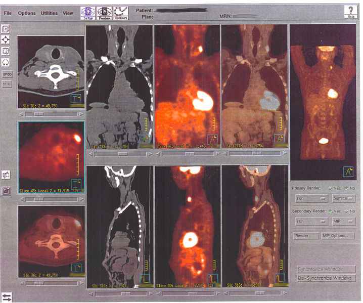
treatment option. PET is often used in conjunction with an MRI
or CT scan through "fusion" to give a full three-dimensional
view of an organ.
PET scan images
38 | Cognitive Psychology and Neuroscience
Behavioral and Neuroscience Methods
fMRI
fMRI (Functional Magnetic Resonance Imaging) is a
technique for determining which parts of the brain are activated
by different types of physical sensation or stimuli such as sight,
sound or the movement of a subject's fingers. The brain
mapping is done by setting up an MRI scanner in a special way
so that the increased blood flow to the activated areas of the
brain shows up on Functional MRI scans. Compared to MRI,
fMRI does not depend on contrast agents although contrast
agents enable far greater detection sensitivity than BOLD
(Blood Oxygenation Level Dependent) signal. Higher BOLD fMRI picture
signal intensities arise from decreases in the concentration of
deoxygenated hemoglobin.
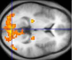
An fMRI experiment usually lasts 1-2 hours. The subject will lie in the magnet and a particular
form of stimulation will be set up and MRI images of the subject's brain are taken. In the first step a
high resolution single scan is taken. This is used later as a background for highlighting the brain areas
which were activated by the stimulus. In the next step a series of low resolution scans are taken over
time, for example, 150 scans, one every 5 seconds. For some of these scans, the stimulus will be
presented, and for some of the scans, the stimulus will be absent. The low resolution brain images in
the two cases can be compared, to see which parts of the brain were activated by the stimulus.
The rest of the analysis is done using a series of tools which correct distortions in the images,
remove the effect of the subject moving their head during the experiment, and compare the low
resolution images taken when the stimulus was off with those taken when it was on. The final statistical
image shows up bright in those parts of the brain which were activated by this experiment. These
activated areas are then shown as coloured blobs on top of the original high resolution scan. This image
can also be rendered in 3D.
fMRI has moderately good spatial resolution. However, the temporal response of the blood supply, which is the basis of fMRI, is poor relative to the electrical signals that define neuronal communication.
Therefore, some research groups are working around this issue by combining fMRI with data collection
techniques such as electroencephalography (EEG) or magnetoencephalography (MEG), which have
much higher temporal resolution but rather poorer spatial resolution.
Electromagnetic Recording Methods
The methods we have mentioned up to now examine the metabolic activity of the brain. But there are also other cases in which one wants to measure electrical activity of the brain or the magnetic fields
produced by the electrical activity. The methods we discussed so far do a great job of identifying where
activity is occurring in the brain. A disadvantage of these methods is that they do not measure brain
activity on a millisecond-by-millisecond basis. This measuring can be done for example by methods as
the single-cell recording or the Electronencephalography (EEG). These methods can measure brain
activity really fast and so they can give a best available temporal resolution.
Wikibooks | 39
Chapter 4
Single cell
When using the single-cell method an electrode is placed into a region of the brain in which we
focus our attention. Now, it is possible for the experimenter to record the electrical output of the cells
that are contacted by the exposed electrode tip. The researchers’ goal is to determine for example if the
cells respond to information from only specific places in the sensory world or from broad regions of
space. Next they want to determine whether the cells are sensitive to input in only one sensory modality
or are multimodal in sensitivity. Furthermore they want to find out if the animal’s attention directed to
a stimulus influence in a cell’s respond.
Single cell studies are not sufficient for studying the human brain, since it is too invasive to be a
common method. Hence, this method is most often used in animals. There are just a few cases in which
the single-cell recording is also applied in humans. People with epilepsy sometimes get removed the
epileptic tissue. A week before surgery electrodes are implanted into the brain or get placed on the
surface of the brain during the surgery to better isolate the source of seizure activity. So using this
method one can decrease the possibility that useful tissues will be removed. Next one can find out
which properties of a stimulus make cells in those regions fire. Due to the limitations of this method in
humans there are other methods which measure electrical activity. Those we are going to discuss next.
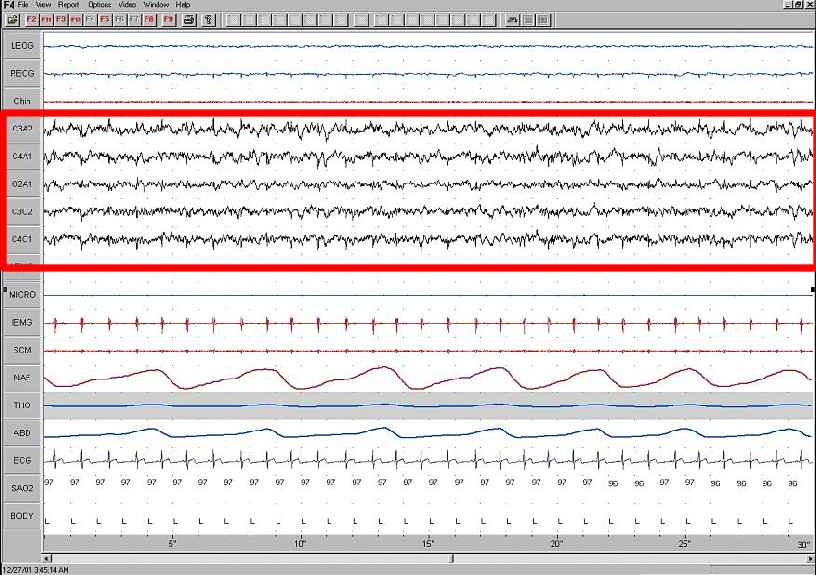
EEG
One of the most famous techniques to study
brain activity is probably the
Electroencephalograhpy (EEG). Most people
might know it as a technique which is used
clinically to detect abberant activity such as that
which accompanies epilepsy and disorders.
In an experimental way this technique is
used to show the brain activity in certain
psychological states, such as alertness or
drowsiness. To measure the brain activity
mental electrodes are placed on the scalp. Each The right placement of the electrodes. ( GFDL - Thekla electrode, also known as lead, acts as its own Helmstedt)
recording site. Next, a reference is needed which provides a
baseline against which the activity at each of the other
electrodes can compared.This electrode must not cover
muscles, because its contractions are induced by electrical
signals Usually this electrode is placed at the mastiod bone
which is located behind the ear.
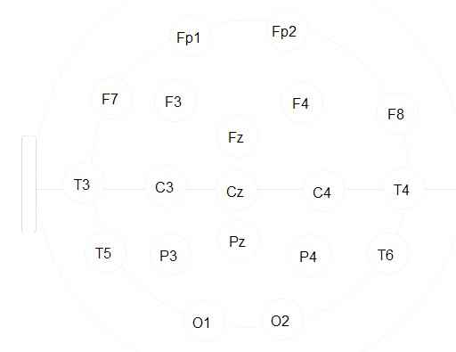
During the EEG electrodes are places like this. Over the
right hemisphere electrodes are labeled with even
numbers.Odd numbers are used for those on the left
hemisphere. Those on the midline are labeled with a z. The
capital letters stands for the location of the
electrode(C=central, F=frontal, Fp= frontal pole, O= occipital,
P= parietal and T= temporal).(see picture)
EEG Stage
40 | Cognitive Psychology and Neuroscience
Behavioral and Neuroscience Methods
After placing each electrode at the right position the electrical potential can be measured. This electrical potential has a particular voltage and furthermore a particular frequency. Accordingly, to a
person’s state the frequency and form of the EEG signal can differ. If a person is awake beta activity
can be recognized, which means that the frequency is relatively fast. Just before someone falls asleep
one can observe alpha activity, which have a slower frequency. The slowest frequencies are called delta
activity, which occur during sleep.
Patients who suffer epilepsy show an increase of the amplitude of firing that can be observed on
the EEG record. In addition EEG can also be used to help answering experimental questions. In the
case of emotion for example, one can see that there is a greater alpha suppression over the right frontal
areas than over the left ones, in the case of depression. One can conclude from this, that depression is
accompanied by greater activation of right frontal regions than of left frontal regions.
ERP
Whereas EEG recordings provide a continuous measure of brain activity, event-related potentials
(ERPs) are recordings which are linked to the occurrence of an event. A presentation of a stimulus
would be such an event. When a stimulus is presented, the electrodes, which are placed on a person’s
scalp, record changes in the brain generated by the thousands of neurons under the electrodes.
By measuring the brain's response to an event we can learn how different types of information are
processed. Representing the word eat or bake for example causes a positive potential at about 200msec.
From this one can conclude, that our brain processes these words 200msec after presenting it. This
positive potential is followed by a negative one at about 400msec. This one is also called N400
(whereas N stands for negative and 400 for the time). So in general one can say that there is a letter P
or N to denote whether the deflection of the electical signal is positive or negative. And a number,
which represent, on average, how many hundreds of milliseconds after stimulus presentation the
component appears.
The event-related- potential shows special interest for researchers, because different components
of the response indicate different aspects of cognitive processing. For example, presenting the
sentences “The cats won’t eat” and “The cat won’t bake”, the N400 response for the word eat is smaller
than for the word bake. From this one can draw the conclusion that our brain needs 400msec to register
information about a word’s meaning. Furthermore, one can figure out where this activity occurs in the
brain, namely if one looks at the position on the scalp of the electrodes that pick up the largest response.
MEG
Magnetoencephalography (MEG) is a related method to the EEG. But instead recording electrical
potentials, it uses magnetic potentials at the scalp to index brain activity. To locate a dipole, the
magnetic field can be used, because the dipole extreme high points of intensity of the magnetic field.
By using devices called SQUIDs (superconducting quantum interference device) MEG can record these
magnetic fields.
MEG is mainly used to localize the source of epileptic activity and to locate primary sensory
cortices. This is helpful because by locating them they can be avoided during neurological intervention.
Wikibooks | 41
Chapter 4
Furthermore, MEG can be used to understand more about the neurophysiology underlying psychiatric
disorders such as schizophrenia. In addition, MEG can also be used to examine a variety of cognitive
processes, such as language, object recognition and spatial processing among others, in people who
were neurologically intact.
MEG has some advantage, because as well known, electrical currents conduct through different
media to different degrees. The electrical current is also carried in different degrees through brain
tissues, cerebral spinal fluid, the skull and the scalp. Magnetic fields instead are not so influenced by
these variations. Another advantage is that the strength of the magnetic field which is recorded can also
tell us information about how deep within the brain the source is located.
However, MEG also has some disadvantages. The magnetic field in the brain is 100 millionth the
size of the earths’ magnetic field. Due to this, shielded rooms, made out of aluminium, are required.
Another disadvantage is that MEG cannot detect activity of cells with certain orientations within the
brain. For example magnetic fields created by cells with long axes radial to the surface will be
invisible.
Techniques for Modulating Brain Activity
Transcranical magnetic stimulation (TMS)
History and procedure
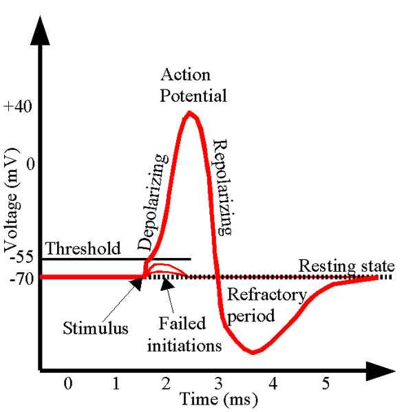
One important technique for modulating brain activity is the so called transcranical magnetic
stimulation, better known as TMS. It is a relatively new technique for inducing small, localized, and
reversible changes in living brain tissue. By using an electromagnet to produce a rapidly fluctuating
magnetic field in the brain, TMS changes the electrical potential in brain tissue, which causes neuronal
Actually, the first modern TMS device was developed by Antony Baker in the year 1985 in
Sheffield after 8 years of research. The field has developed rapidly since than with many researchers
using TMS in order to study a variety of brain functions. It
has been used, e.g., to block the perception of visual stimuli,
in order to cause speech arrest, and to delay the onset of
voluntary movements. The clinical effects of TMS have also
been investigated in certain neuropsychiatric conditions.
Several investigators have suggested the possible efficacy of
TMS as a treatment for depression in human trials and animal
models. Because of findings such as these, TMS has been
considered as a possible alternative to antidepressant
medication. (1, 2)
Mechanisms
Although TMS is able to influence many brain functions, Action potential ( GFDL - Chris 73)